. 2008 Jan 9;27(1):277-89.
doi: 10.1038/sj.emboj.7601936. Epub 2007 Dec 6.

Dimerization of FIR upon FUSE DNA binding suggests a mechanism of c-myc inhibition

Affiliations

Dimerization of FIR upon FUSE DNA binding suggests a mechanism of c-myc inhibition

Gregg V Crichlow et al. EMBO J. .

Abstract

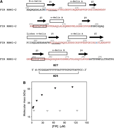
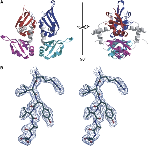
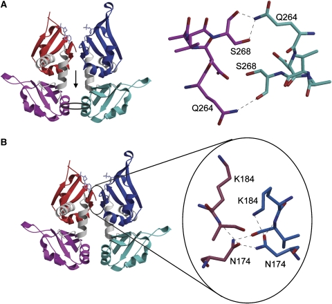
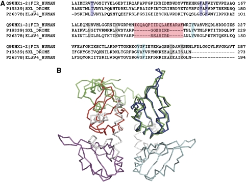
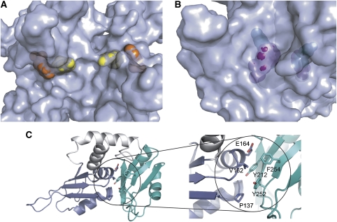
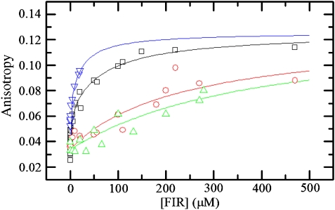
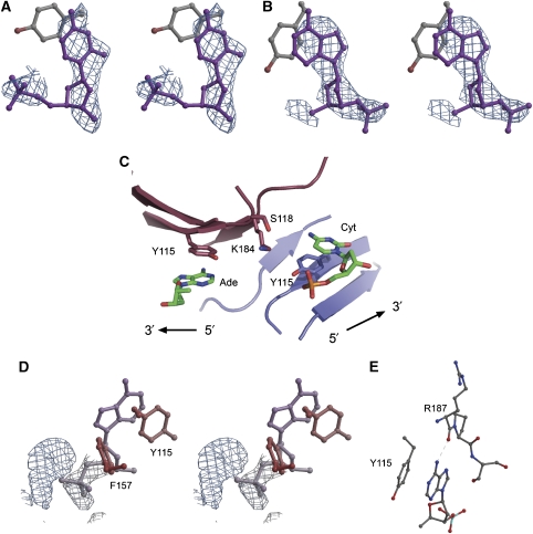
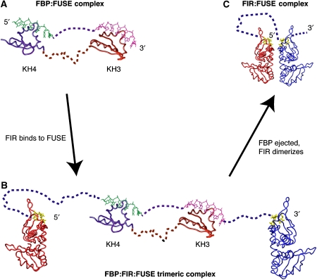
c-myc is essential for cell homeostasis and growth but lethal if improperly regulated. Transcription of this oncogene is governed by the counterbalancing forces of two proteins on TFIIH--the FUSE binding protein (FBP) and the FBP-interacting repressor (FIR). FBP and FIR recognize single-stranded DNA upstream of the P1 promoter, known as FUSE, and influence transcription by oppositely regulating TFIIH at the promoter site. Size exclusion chromatography coupled with light scattering reveals that an FIR dimer binds one molecule of single-stranded DNA. The crystal structure confirms that FIR binds FUSE as a dimer, and only the N-terminal RRM domain participates in nucleic acid recognition. Site-directed mutations of conserved residues in the first RRM domain reduce FIR's affinity for FUSE, while analogous mutations in the second RRM domain either destabilize the protein or have no effect on DNA binding. Oppositely oriented DNA on parallel binding sites of the FIR dimer results in spooling of a single strand of bound DNA, and suggests a mechanism for c-myc transcriptional control.


Figures

Figure 1
Figure 1
Sequence and solution characterization of FIR1+2:FUSE complex (A) Sequence of protein and DNA constructs used in this study. The sequence and secondary structure of FIR1+2 corresponding to human FIR residues 101–299 is shown. The corresponding sequences of PUF60 and Siah-BP are identical to FIR RRM1+2. The RRM domains are colored in red, and residues mutated from Cys are colored in blue. The sequence of each RNP1 is boxed, and the sequence of each RNP2 is underlined in each RRM domain. (B) Oligomeric state of FIR–DNA complex from SEC-UV/LS/RI analysis. Weight-average MWs determined from SEC-UV/LS/RI analyses are plotted as filled triangles for FIR–DNA complex and open triangles for FIR protein alone. Averages and standard deviations were calculated from 25 MW determinations for the top 0.2 ml of the eluting peaks for which the concentration is within 5% of the plotted value.
Figure 2
Figure 2
Structure of FIR1+2 bound to FUSE DNA. (A) Ribbon representation showing the domain organization. RRM domains 1 and 2 of subunit A are in red and magenta, respectively. RRM domains 1 and 2 of subunit B are in blue and cyan, respectively. DNA is shown in purple. Residues 147–150 and 179 of subunit A, where electron density is poor, are not included in the figure. The view on the right is rotated 90° from that on the left. The two-fold axis relating the two subunits is in the plane of the page in both views. (B) Stereo-view of 1.0σ2FoFc electron density of a portion of the protein from the β1-strand of RRM2 (subunit B). This figure and parts of Figure 7 were prepared using Molscript and Bobscript (Kraulis, 1991; Merritt and Bacon, 1997).
Figure 3
Figure 3
Intersubunit interactions in the FIR–HJ25 complex. (A) The intersubunit interactions involving the RRM2 domains are shown (right) looking toward the surface in the direction of the arrow in the left panel. (B) Hydrogen bonding network involving RRM1 in the subunit interface, showing the participation of Asn 174 and Lys 184. The orientation is with the DNA-binding region toward the top. This figure was prepared using Molscript.
Figure 4
Figure 4
Comparison of FIR with monomeric tandem RRM domain proteins. (A) Sequence alignment showing the RRM domains in FIR1+2 and the corresponding amino acids in the D. melanogaster protein sex-lethal and in the human HuD antigen (also known as ELAV-like protein 4). Highlighted in purple are the RNP1 and two central nucleic –acid-interacting residues of RRM1, and in cyan those of RRM2. In gray are the residues corresponding to those in FIR that form hydrogen bonds across the subunit interface. In pink is the linker region between the two RRM domains. (B) Structural overlay of Drosophila sex-lethal (PDB entry 1B7F) on the FIR1+2 dimer. The overlay was performed by least-squares superposition of α-carbons in the first RRM domain of sex-lethal onto the corresponding α-carbons of the first RRM domain of a subunit of FIR. The nucleic acid from both structures is omitted for clarity. This, as well as Figures 5 and 7C, was prepared using PyMol (DeLano, 2002).
Figure 5
Figure 5
RRM–interdomain contacts. (A, B). Surface representations of FIR1+2 contrasting the surface exposure of DNA-recognition residues of the N-terminal RRM domain (RRM 1) to the relative inaccessibility of corresponding residues of RRM 2. In panel A, Tyr115 (yellow) and Phe157 (orange) are both solvent exposed. In panel B, Tyr212 (green) is completely buried and inaccessible to solvent, and Phe254 (purple) is mostly buried. (C) Cartoon and stick representation of the hydrophobic interactions between the two domains. Aromatic residues in the putative nucleic acid-binding site of RRM2 are involved in interdomain hydrophobic contacts. The RRM1 domain is colored blue, and the RRM2 domain is colored cyan, as in Figure 2.
Figure 6
Figure 6
Equilibrium binding affinities of wildtype and mutant FIR determined by fluorescence anisotropy. Steady-state anisotropy of 3′-fluorescein-labeled H27 is plotted as a function of FIR concentration for FIR1+2 wildtype (black squares), Y115L (red circles), F157L (green triangles) and F254L (blue upside-down triangles). Lines depict the best fits of [FIR] dependence of the anisotropy data to equation 3. The equilibrium binding affinities obtained from the best fits of [FIR] dependence of the anisotropy data to equation 3 are presented in Table II.
Figure 7
Figure 7
(A) Nucleotide stacked against Tyr115 in subunit B, with 1.0σ2FoFc electron density, calculated by omitting the DNA from the model. (B) 2.5σFoFc electron density, calculated by omitting the DNA from the model. The adenine stacking against Tyr115 in subunit A is shown. (C) A view of the DNA-binding site centered on the dimeric interface, showing the base stacking of Tyr115 of each subunit with nucleotide. Subunit A is depicted in maroon and subunit B in blue, similar to Figure 2A. (D) View of unmodelled 1.0σ2FoFc electron density corresponding to DNA near Phe157. Shown is subunit B, but unmodelled density is also present in subunit A. (E) Hydrogen bond between adenine and Arg187.
Figure 8
Figure 8
Model of progression in FBP and FIR interaction with FUSE during c-myc transcription. FUSE becomes single stranded upon the initiation of c-myc transcription and is recognized by FBP. Shortly thereafter, FIR binds the FBP/FUSE complex, forming a trimeric complex. Finally, FBP is ejected and FIR forms a long-lived stable complex with FUSE. (A) Molecular dynamic studies of the third and fourth KH domains of FBP bound to FUSE show that FBP binds FUSE as an elongated molecule in which the DNA is flexible but linear (from Braddock et al, 2002a). (B, C) SEC/LS/RI/UV measurements demonstrate that FIR binds FUSE as a monomer at low concentrations, but dimerizes at higher concentrations in the presence of ssDNA. FIR dimerization would change the topology of FUSE, helping to pry the C-terminal activation domain of FBP of TFIIH and facilitate the ejecting FBP from FUSE.

Similar articles

Cited by

References

    1. Allain FH, Gilbert DE, Bouvet P, Feigon J (2000) Solution structure of the two N-terminal RNA-binding domains of nucleolin and NMR study of the interaction with its RNA target. J Mol Biol 303: 227–241 - PubMed
    1. Amrute SB, Abdul-Manan Z, Pandey V, Williams KR, Modak MJ (1994) Purification and nucleic acid binding properties of a fragment of type C1/C2 heterogeneous nuclear ribonucleoprotein from thymic nuclear extracts. Biochemistry 33: 8282–8291 - PubMed
    1. Auweter SD, Oberstrass FC, Allain FH (2006) Sequence-specific binding of single-stranded RNA: is there a code for recognition? Nucleic Acids Res 34: 4943–4959 - PMC - PubMed
    1. Bae E, Reiter NJ, Bingman CA, Kwan SS, Lee D, Phillips GN Jr, Butcher SE, Brow DA (2007) Structure and interactions of the first three RNA recognition motifs of splicing factor prp24. J Mol Biol 367: 1447–1458 - PMC - PubMed
    1. Braddock DT, Baber JL, Levens D, Clore GM (2002a) Molecular basis of sequence-specific single-stranded DNA recognition by KH domains: solution structure of a complex between hnRNP KKH3 and single-stranded DNA. EMBO J 21: 3476–3485 - PMC - PubMed

Publication types

MeSH terms

Associated data