

doi: 10.1364/ol.32.003450.
Retinal flow cytometer
Affiliations
- PMID: 18059963
- PMCID: PMC2794483
- DOI: 10.1364/ol.32.003450
Item in Clipboard
Retinal flow cytometer
Opt Lett.
.
Abstract
The in vivo flow cytometer is an instrument capable of continuous, real-time monitoring of fluorescently labeled cells in the circulation without the need to draw blood samples. However, the original system probes a single vessel in the mouse ear; the small sample volume limits the sensitivity of the technique. We describe an in vivo retinal flow cytometer that simultaneously probes five artery-vein pairs in the mouse eye by circularly scanning a small laser spot rapidly around the optic nerve head. We demonstrate that the retinal flow cytometer detects about five times more cells per minute than the original in vivo flow cytometer does in the ear.
Figures
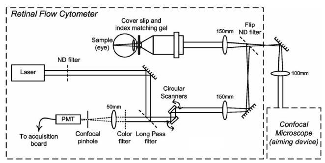
Schematic of the retinal flow cytometer setup. Two resonant scanners create the circular scan on the retina. Fluorescence is detected through a long-pass and a bandpass filter. A confocal pinhole in front of the PMT rejects the out-of-focus signal.
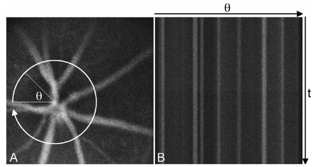
Confocal fluorescence image of mouse retinal vessels visualized with the fluorescent dye Evans Blue, A, with a cartoon of the circular retinal flow cytometer scan. Evans Blue has similar excitation and emission characteristics as DiD. Consecutive circular scans are mapped to straight horizontal lines in the retinal flow cytometer file, B. Thus retinal vessels appear as vertical, fluorescent structures.
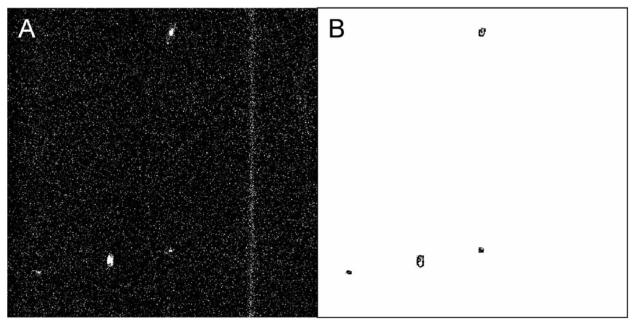
Typical frame of retinal flow cytometer raw file from DiD-labeled lymphocytes, A, and the same frame analyzed by ImageJ, B. The four cells in the raw file mark the position of three blood vessels (oriented vertically; compare Fig. 2). The long streak is a single cell that is moving very slowly in a capillary. This location was rejected from the analysis throughout the data set since it is not a major retinal vessel. The other four cells are correctly counted and outlined.
References
-
- Georgakoudi I, Solban N, Novak J, Rice WL, Wei X, Hasan T, Lin CP. Cancer Res. 2004;64:5044. - PubMed
Publication types
MeSH terms
Grants and funding
LinkOut - more resources
Full Text Sources
Other Literature Sources
