. 2007 Dec;117(12):3753-64.
doi: 10.1172/JCI32481.

CP-31398 restores mutant p53 tumor suppressor function and inhibits UVB-induced skin carcinogenesis in mice

Affiliations

CP-31398 restores mutant p53 tumor suppressor function and inhibits UVB-induced skin carcinogenesis in mice

Xiuwei Tang et al. J Clin Invest. 2007 Dec.

Abstract

Mutations in the tumor suppressor p53 are detectable in over 50% of all human malignancies. Mutant p53 protein is incapable of transactivating its downstream target genes that are required for DNA repair and apoptosis. Chronic exposure to UVB induces p53 mutations and is carcinogenic in both murine and human skin. CP-31398, a styrylquinazoline compound, restores the tumor suppressor functions of mutant forms of p53 in tumor cells. However, its effectiveness in vivo remains unclear. Here, we demonstrate that CP-31398 blocked UVB-induced skin carcinogenesis and was associated with increases in p53, p21, and BclXs. CP-31398 downregulated Bcl2, proliferating nuclear cell antigen, and cyclin D1. Activation of caspase-3 and cleavage of poly (ADP-ribose) polymerase also occurred in both tumor and perilesional skin following treatment. CP-31398 induced the expression of p53-dependent target proteins, and this was followed by apoptosis in UVB-irradiated wild-type mice but not in their p53-deficient littermates. Similar effects were observed in human skin carcinoma A431 cells expressing mutant p53. In addition, CP-31398 induced mitochondrial translocation of p53, leading to changes in mitochondrial membrane permeability pore transition (MPT) and consequent cytochrome c release in these cells. Blocking MPT diminished p53 translocation and apoptosis. These studies indicate that reconstituting p53 tumor suppressor functions in vivo by small molecular weight compounds may block the pathogenesis and progression of skin cancer.


Figures

Figure 1
Figure 1. Effects of CP-31398 treatment on the expression of p53 target, cell-cycle regulatory, and apoptosis-related proteins in the skin of mice chronically irradiated with UVB.
(A) Western blot showing expression of p53 and its downstream target gene p21CIP1 in UVB-irradiated skin of SKH-1 mice treated with CP-31398. (B) The expression of proapoptotic BclXs, antiapoptotic Bcl2, and cell-cycle regulatory cyclin D1 in UVB-irradiated skin of SKH-1 mice following CP-31398 treatment. The treatment protocol and other experimental details are provided in Methods.
Figure 2
Figure 2. Effects of CP-31398 treatment on the growth of UVB-induced skin tumors in SKH-1 mice.
(A) Drawing showing experimental protocol design. CP-31398 administered i.p. reduces growth of UVB-induced skin tumors in SKH-1 mice in terms of tumor number (lower left panel) and tumor volume (lower right panel). (B) Effect of CP-31398 on the expression of p53 and its downstream target genes PUMA-α and mdm-2 in perilesional skin and tumors. (C) Effect of CP-31398 on the expression of cyclins A, B1, D1, and E in perilesional skin and tumors. (D) PARP cleavage and expression of antiapoptotic Bcl2 in perilesional skin and tumors following CP-31398 treatment. (E) Immunohistochemical analyses showing expression of Bcl2 and Bax in UVB-irradiated skin and UVB-induced SCC following CP-31398 treatment. Arrows indicate Bax- or BCL2-expressing cells. Treatment protocol and other experimental details are provided in Methods. P values represent significance when compared with vehicle-treated control at corresponding time point.
Figure 3
Figure 3. Chemopreventive effects of CP-31398 on UVB-induced skin photocarcinogenesis.
(A) Drawing showing experimental protocol design. Topical CP-31398 reduces UVB-induced cutaneous tumor growth in terms of tumor number (lower left panel) and tumor size (lower right panel). (B) Picture of representative mice showing effects of CP-31398 on UVB-induced skin tumor development. (C) Expression of cyclins B1, D1, and A in UVB-induced skin tumors of SKH-1 mice following CP-31398 treatment. Treatment protocol and other experimental details are provided in Methods. P values represent significance when compared with vehicle-treated control at corresponding time points.
Figure 4
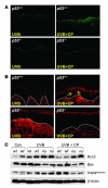
Figure 4. Effects of CP-31398 treatment on the induction of apoptosis in the skin of UVB-irradiated p53+/+/SKH-1 and p53–/–/SKH-1 mice.
(A) TUNEL staining showing CP-31398–induced apoptosis in UVB-irradiated skin of p53+/+/SKH-1 and p53–/–/SKH-1 hairless littermates. (B) Immunostaining for Smac/Diablo in epidermis of UVB-irradiated skin of p53+/+/SKH-1 and p53–/–/SKH-1 hairless littermates following CP-31398 treatment. Arrows show release of Smac from mitochondria to cytoplasm. (C) Western blot showing the expression of Bcl2, Bax, and PARP cleavage in UVB-irradiated skin of p53+/+/SKH-1 and p53–/–/SKH-1 hairless littermates. Treatment protocol and other experimental details are provided in Methods.
Figure 5
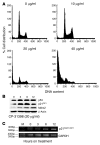
Figure 5. Effects of CP-31398 treatment on cell-cycle progression in human epidermoid carcinoma A431 cells.
(A) CP-31398 induces cell-cycle arrest in A431 cells. (B) Western blot showing the expression of p53 and its downstream target genes p21CIP1 and mdm2 in A431 cells treated with CP-31398. (C) RT-PCR showing mRNA levels of p21CIP1 in CP-31398–treated A431 cells. A431 cells were treated with PBS (control) or various concentrations of CP-31398 for different time intervals as indicated in the figure. For Western blot analysis, 100 μg protein was loaded in each well. Each experiment was repeated at least 3 times.
Figure 6
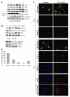
Figure 6. Effects of CP-31398 treatment on the induction of apoptosis in neonatal human epidermal keratinocytes and human epidermoid carcinoma A431 cells.
(A) Western blot showing the expression of apoptosis proteins. (B) Western blot showing the expression of cell-cycle regulatory proteins in A431 cells following CP-31398 treatment. (C) Effect of CP-31398 on the induction of apoptosis and release of mitochondrial proteins in NHEKs carrying wild-type p53 and A431 cells carrying mutant p53. (D) Effect of CP-31398 on the changes in mitochondrial membrane potential in A431 cells. A431 cells were treated with PBS (control) or various concentrations of CP-31398 as indicated in the figure for different time intervals. For Western blot analysis, 100 μg protein was loaded in each well. Each experiment was repeated at least 3 times. For assaying mitochondrial membrane potential, A431 cells were treated with PBS (control) or 10, 20, and 40 μg/ml of CP-31398 for 10 minutes and stained with JC-1 dye. Carbonyl cyanide 3-chlorophenylhydrazone (CCCP) was used as positive control. Error bars represent mean ± SD of triplicate samples.
Figure 7
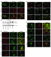
Figure 7. Effects of CP-31398 on the translocation of p53 to mitochondria, alternations in MPT potential, and release of cytochrome c.
(A) Immunofluorescence staining showing colocalization of p53 with MitoTracker, which stains for mitochondria. (B) Western blots of immunocoprecipitates of p53 and mitochondrial MnSOD from mitochondrial protein extracts prepared from CP-31398–treated, vehicle-treated, and untreated A431 cells. (C) Immunofluorescence staining showing cyclosporin A blocks CP-31398–induced migration of p53 to mitochondria of A431 cells. (D) Immunofluorescence staining (using MitoCapture) showing cyclosporin A blocks CP-31398–induced apoptosis in A431 cells. Arrows indicate localization of p53 in mitochondria in A and C, whereas in D, arrows show live cells with red fluorescence and green fluorescence representing cells undergoing apoptosis. (E) Immunofluorescence staining showing cyclosporin A blocks CP-31398–induced release of cytochrome c in A431 cells. A431 cells were treated with PBS (control) or various concentrations of CP-31398 for different time intervals as described in Methods or indicated in the figure. Each experiment was repeated at least 3 times. For immunoprecipitation followed by Western blot analysis, 300 μg protein was used.
Figure 8
Figure 8. Cascade of events occurring during CP-31398–mediated activation of mutant p53 to wild-type function.
CP-31398 interacts with mutant p53 and restores its wild-type function, including transcriptional activation of its target genes, such as p21, mdm2, puma, bax, and apaf-1. The enhanced transcription and translation of these genes led to cell-cycle arrest followed by apoptosis. The mechanism of apoptosis induction involves translocation of p53 to mitochondria, and that alters MPT, which is followed by the release of mitochondria-localized cytochrome c and Smac/Diablo. Smac/Diablo release leads to blockade of the survivin function and results in the activation of caspase-3. Activation of this pathway triggers apoptosis in tumor cells carrying mutant p53, ultimately leading to tumor ablation.

Comment in

Similar articles

Cited by

References

    1. Vogelstein B., Kinzler K.W. Cancer genes and the pathways they control. Nat. Med. 2004;10:789–799. - PubMed
    1. Vogelstein B., Lane D., Levine A.J. Surfing the p53 network. Nature. 2000;408:307–310. - PubMed
    1. van Kranen H.J., de Gruijl F.R. Mutations in cancer genes of UV-induced skin tumors of hairless mice. J. Epidemiol. 1999;9:S58–S65. - PubMed
    1. Phan T.A., Halliday G.M., Barnetson R.S., Damian D.L. Spectral and dose dependence of ultraviolet radiation-induced immunosuppression. Front. Biosci. 2006;11:394–411. - PubMed
    1. D’Errico M., Lemma T., Calcagnile A., Santis L.P.D., Dogliotti E. Cell type and DNA damage specific response of human skin cells to environmental agents. Mutat. Res. 2007;614:37–47. - PubMed

Publication types

MeSH terms