. 2007 Dec;117(12):3774-85.
doi: 10.1172/JCI32311.

Microvascular destruction identifies murine allografts that cannot be rescued from airway fibrosis

Affiliations

Microvascular destruction identifies murine allografts that cannot be rescued from airway fibrosis

Ashok N Babu et al. J Clin Invest. 2007 Dec.

Abstract

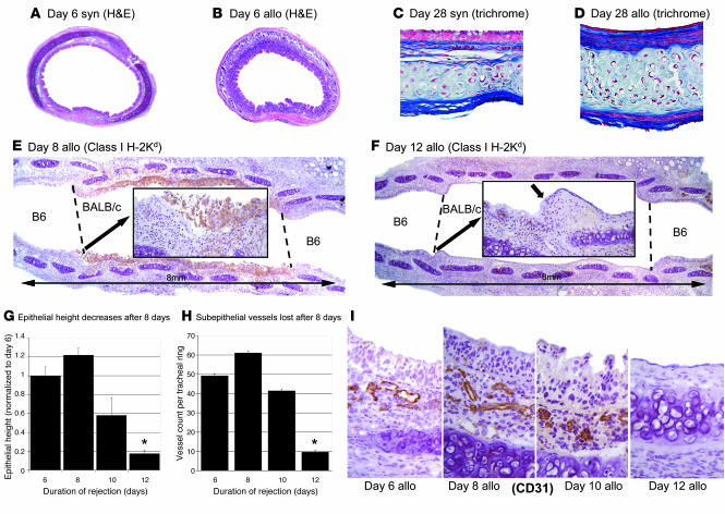
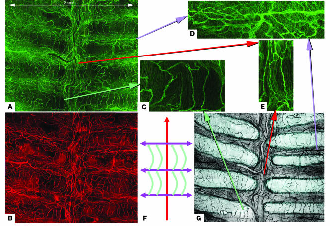
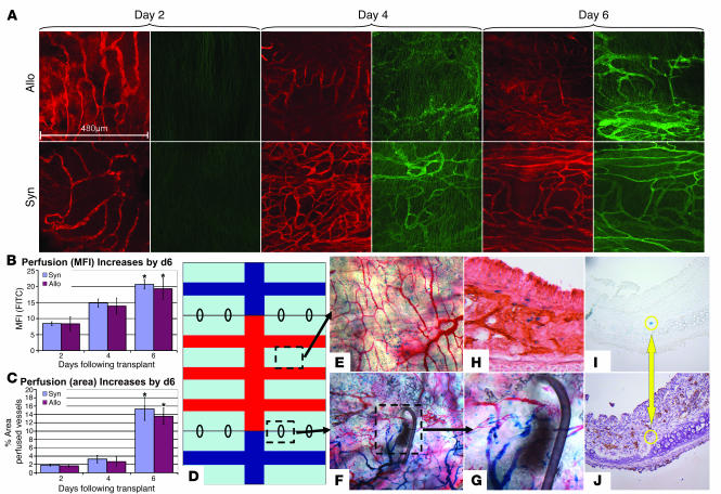
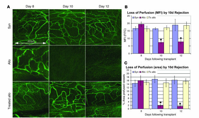
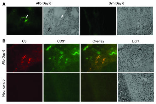
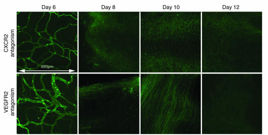
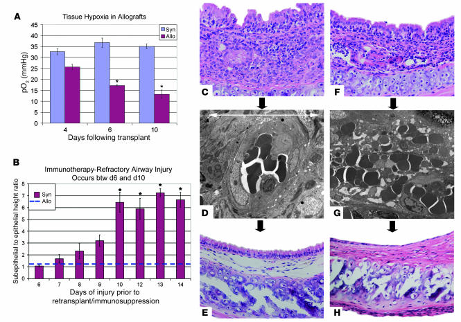
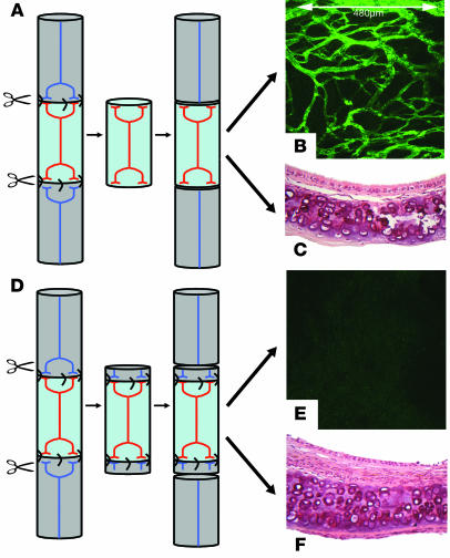
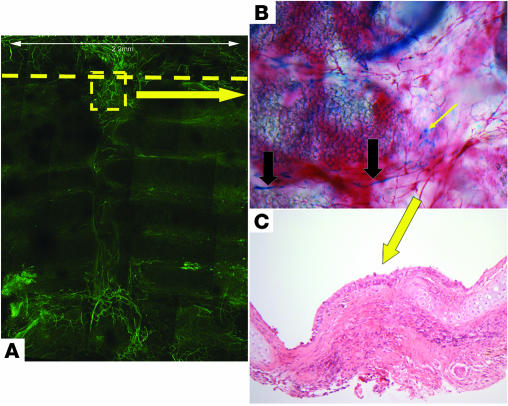
Small airway fibrosis (bronchiolitis obliterans syndrome) is the primary obstacle to long-term survival following lung transplantation. Here, we show the importance of functional microvasculature in the prevention of epithelial loss and fibrosis due to rejection and for the first time, relate allograft microvascular injury and loss of tissue perfusion to immunotherapy-resistant rejection. To explore the role of alloimmune rejection and airway ischemia in the development of fibroproliferation, we used a murine orthotopic tracheal transplant model. We determined that transplants were reperfused by connection of recipient vessels to donor vessels at the surgical anastomosis site. Microcirculation through the newly formed vascular anastomoses appeared partially dependent on VEGFR2 and CXCR2 pathways. In the absence of immunosuppression, the microvasculature in rejecting allografts exhibited vascular complement deposition, diminished endothelial CD31 expression, and absent perfusion prior to the onset of fibroproliferation. Rejecting grafts with extensive endothelial cell injury were refractory to immunotherapy. After early microvascular loss, neovascularization was eventually observed in the membranous trachea, indicating a reestablishment of graft perfusion in established fibrosis. One implication of this study is that bronchial artery revascularization at the time of lung transplantation may decrease the risk of subsequent airway fibrosis.


Figures

Figure 1
Figure 1. Loss of epithelium and subepithelial vessels after 8 days of acute rejection.
(A) Radial section of syngeneic (B6→B6) tracheal graft 6 days after transplant demonstrating normal columnar epithelium. (B) Allogeneic (BALB/c→B6) graft with massive inflammatory cell infiltration of the subepithelium and epithelium 6 days following transplant. (C and D) Masson’s trichrome stain demonstrates subepithelial fibrosis and epithelial changes at 28 days in allografts as compared with normal histology in syngeneic grafts. (E) Coronal section of tracheal allograft at 8 days (n = 4) stained for MHC class I H-2Kd (BALB/c) demonstrates donor-type columnar epithelium in the graft without staining of recipient epithelium. (F) Coronal section of allograft at 12 days immunostained for MHC class I H-2Kd demonstrates replacement of columnar epithelium with flattened epithelium (short arrow). (G) Morphometric analysis of epithelial height demonstrates epithelial height loss between 8 days and 12 days. (H and I) Radial sections of tracheal allografts and corresponding vessel counts demonstrate loss of subepithelial vessels after 8 days of rejection. *P < 0.01 versus all other groups; n = 4–5 for all groups. allo, allogeneic; syn, syngeneic. Original magnification, ×4 (A, B, H, and I); ×20 (C and D).
Figure 2
Figure 2. Tracheal microvasculature is organized based on cartilaginous anatomy.
(A) Following i.v. injection of FITC-conjugated (green) tomato lectin while still alive, naive animals were sacrificed and tracheae whole mounted to visualize the tracheal vasculature. (B) These same tracheae underwent immunostaining for CD31 (endothelial cell antigen) with a Cy3 (red) secondary antibody after excision to identify all vessels regardless of perfusion status. (C) Magnified image of vessels originating from the intercartilaginous vessels (D) that span the cartilage rings. (D) Transverse vessels carrying blood away from the midline to the intercartilaginous trachea to supply cartilage-spanning vessels in C. (E) Axial direction vessels in the midline membranous trachea, which appear to be major blood highway for trachea. (F) Schematic depiction of tracheal blood flow, which is color-coordinated with previous cutout images. (G) Cartoon version of image A demonstrating cartilage rings in light blue and intercartilaginous or membranous regions in dark gray. Original magnification, ×20.
Figure 3
Figure 3. Both allogeneic and syngeneic grafts reperfuse by day 6 via connections of recipient vessels to the preexisting donor vascular network at the anastomosis.
(A) At day 2, both allografts and syngrafts have a CD31+ vascular network (red) but no perfusion. Reperfusion of both allografts and syngeneic grafts begins at day 4 and is complete by day 6. (B and C) Mean fluorescent intensity and percent area of FITC+ vessels. *P < 0.05 versus both groups at day 2 and day 4. (D) Schematic of transplanted trachea. Black ovals represent sutures at anastomosis site. Light blue represents the cartilage rings. Red colors the membranous trachea in the transplanted graft, and blue represents that of the recipient ends. (EG) Vessel origin experiment was conducted in allografts (BALB/c→FVB [Tie2 / β-galactosidase]) and syngrafts (FVB→FVB [Tie2 / β-galactosidase]). Prior to tissue harvest, these animals were perfused with red india ink via the aorta to opacify perfused vessels in red. (E) Graft vessels are exclusively red in color (i.e., donor origin). (F) Cutout at anastomosis with suture demonstrating recipient vessels, which connect to donor vessels. Blue-staining cells are noted outside of vessels. (G) Higher magnification at anastomosis. (H) Double stain for β-galactosidase and CD31 on axial sectioning of day 6 allograft demonstrating that the blue dots are neither part of the CD31 vascular network nor express CD31. (I and J) Serial sections of day 6 allograft independently stained for β-galactosidase (I) and CD31 (J) to demonstrate that β-galactosidase–expressing cells do not express CD31 (n = 3–4 for all groups). Original magnification, ×20 (E, F, I, and J); ×40 (G and H).
Figure 4
Figure 4. Vascular perfusion is lost by day 10 in untreated allografts and is preserved in syngrafts and immunosuppressed allografts (n = 3 per box).
(A) Normal vascular perfusion is maintained at day 8, day 10, and day 12 in syngrafts. However, there is complete loss of perfusion in allografts at day 10 and day 12. Perfusion of the vessels is completely preserved by immunosuppression at both day 10 and day 12. Treated allo, allograft treated with immunosuppression (anti–LFA-1 + anti-CD40L). (B) FITC–mean fluorescent intensity was measured as an index of blood flow, confirming the loss of blood flow at day 10 and day 12 in untreated allografts. MFI, mean fluorescent intensity (C) Using a mask based on FITC threshold to mark blood vessels, the percentage area of perfused vessels was assessed to confirm the histologic data. *P < 0.05 versus all other groups.
Figure 5
Figure 5. C3 deposition on endothelium of rejecting allografts.
(A) Immunofluorescent (FITC; green) staining for C3 demonstrates positivity in day 6 allografts and none in day 6 syngeneic grafts. (B) Double staining for C3 (Cy3; red) and CD31 (FITC; green) in day 6 allografts demonstrates colocalization (appearing orange) of C3 staining to the vascular endothelium. Original magnification, ×40.
Figure 6
Figure 6. VEGFR2 and CXCR2 antagonism lead to early loss of perfusion.
Animals administered a CXCR2 antagonist (antileukinate; n = 3/time point) or a VEGFR2 antagonist (SU5416; n = 3/time point) were sacrificed at days 6, 8, 10, and 12, and tracheal perfusion was studied by FITC-conjugated lectin perfusion staining. Interestingly, these therapies had no effect on the revascularization occurring by 6 day. However, the loss of perfusion occurred by day 8 rather than by day 10 as in untreated allografts.
Figure 7
Figure 7. Injured and nonperfused endothelium is a marker for injury severity that cannot be rescued by immunosuppression.
(A) Tissue oximetry was performed using a microprobe to detect tracheal epithelial pO2. Syngrafts maintain consistent tissue oxygen content, whereas allografts develop relative tissue hypoxia by 10 day. (B) Allografts (BALB/c→B6) were allowed to undergo varying durations of rejection, at which point they were retransplanted to naive immunosuppressed B6 animals for an additional 28 days. H&E-stained axial sections were used to measure the ratio of subepithelial height to epithelial height. This measure has been validated previously as correlating strongly with chronic rejection histologic scoring (9). Syngeneic grafts that were retransplanted maintained a normal ratio of approximately 1. Rejection durations of 10 day or longer resulted in severe airway remodeling despite rescue therapy (n = 5/group). *P < 0.05 versus all other groups. (C) Axial section of allograft following 6 days rejection. (D) EM demonstrating normal appearance of vascular endothelium in an allograft following 6 days rejection. (E) Axial section of allograft that underwent 6 days of rejection prior to retransplantation to a naive immunosuppressed animal demonstrating rescue with normal histology. (F) Axial section of allograft following 10 days rejection. (G) EM demonstrating abnormal appearance of vascular endothelium with red blood cell extravasation in an allograft following 10 days rejection. (H) Axial section of allograft that underwent 10 days of rejection prior to retransplantation to a naive immunosuppressed animal demonstrating lack of rescue due to flattened epithelium and subepithelial fibrosis. Original magnification, ×20 (C, E, F, and H); ×1,100 (G).
Figure 8
Figure 8. Additional transection of blood vessels during retransplantation affects perfusion and leads to fibrosis.
As shown in Figure 7B, BALB/c→B6 allografts having undergone 6 days of rejection can be rescued from fibrosis by retransplantation into a naive B6 immunosuppressed hosts. (A) The standard retransplantation technique involves cutting only the graft from the original recipient at the original anastomosis site and retransplanting it. (B) Ten days following retransplant into naive immunosuppressed host, there is normal perfusion throughout the graft. (C) By 28 days, histology is normal with no fibrosis and well-differentiated epithelium. (D) To determine whether manipulating the blood flow to a rescuable airway could affect end outcome, we divided the day 6 allograft through the recipient ends, creating an additional transection of vessels. (E) At 10 day following retransplant, there was no perfusion, in stark contrast to B. (F) By day 28, this airway had progressed to epithelial flattening and subepithelial fibrosis. Original magnification, ×20 (C, E, and F).
Figure 9
Figure 9. Neovascularization of membranous trachea by day 28 involves recipient-derived endothelium and supports a well-differentiated epithelium.
(A) Neovascularization is evident by perfusion lectin staining in the membranous and intercartilaginous portions of the trachea following 28 days of rejection (n = 4). The dashed line indicates the anastomosis site separating the recipient and donor. (B) Use of FVB (Tie2/β-galactosidase) recipients (n = 3) demonstrated that by day 28, new vessels were composed partially of recipient-derived endothelium (large black arrows), and β-galactosidase expressing cells (small yellow arrow) appeared to still be present in the allograft in the proximity of vessels. (C) Five of 8 specimens studied demonstrate columnar epithelium only overlying the membranous portion of the trachea following 28 days of rejection, suggesting that the blood supply in this region may be required to support this epithelial phenotype. Original magnification, ×20 (B and C).

Comment in

  • Every allograft needs a silver lining.
    Contreras AG, Briscoe DM. Contreras AG, et al. J Clin Invest. 2007 Dec;117(12):3645-8. doi: 10.1172/JCI34238. J Clin Invest. 2007. PMID: 18060023 Free PMC article.

Similar articles

Cited by

References

    1. Trulock E.P., et al. Registry of the International Society for Heart and Lung Transplantation: twenty-third official adult lung and heart-lung transplantation report — 2006. J. Heart Lung Transplant. 2006;25:880–892. - PubMed
    1. Yousem S.A., Burke C.M., Billingham M.E. Pathologic pulmonary alterations in long-term human heart-lung transplantation. Hum. Pathol. 1985;16:911–923. - PubMed
    1. Sharples L.D., McNeil K., Stewart S., Wallwork J. Risk factors for bronchiolitis obliterans: a systematic review of recent publications. . J. Heart Lung Transplant. 2002;21:271–281. - PubMed
    1. Luckraz H., et al. Microvascular changes in small airways predispose to obliterative bronchiolitis after lung transplantation. J. Heart Lung Transplant. 2004;23:527–531. - PubMed
    1. Luckraz H., et al. Is obliterative bronchiolitis in lung transplantation associated with microvascular damage to small airways? Ann. Thorac. Surg. 2006;82:1212–1218. - PubMed

Publication types

MeSH terms

Substances