. 2006 Dec 6;6(5):261-281.
doi: 10.1016/j.cnr.2006.09.006.

Neuroinflammation, Oxidative Stress and the Pathogenesis of Parkinson's Disease

Neuroinflammation, Oxidative Stress and the Pathogenesis of Parkinson's Disease

R Lee Mosley et al. Clin Neurosci Res. .

Abstract

Neuroinflammatory processes play a significant role in the pathogenesis of Parkinson's disease (PD). Epidemiologic, animal, human, and therapeutic studies all support the presence of an neuroinflammatory cascade in disease. This is highlighted by the neurotoxic potential of microglia . In steady state, microglia serve to protect the nervous system by acting as debris scavengers, killers of microbial pathogens, and regulators of innate and adaptive immune responses. In neurodegenerative diseases, activated microglia affect neuronal injury and death through production of glutamate, pro-inflammatory factors, reactive oxygen species, quinolinic acid amongst others and by mobilization of adaptive immune responses and cell chemotaxis leading to transendothelial migration of immunocytes across the blood-brain barrier and perpetuation of neural damage. As disease progresses, inflammatory secretions engage neighboring glial cells, including astrocytes and endothelial cells, resulting in a vicious cycle of autocrine and paracrine amplification of inflammation perpetuating tissue injury. Such pathogenic processes contribute to neurodegeneration in PD. Research from others and our own laboratories seek to harness such inflammatory processes with the singular goal of developing therapeutic interventions that positively affect the tempo and progression of human disease.


Figures
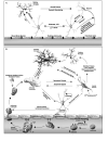
Figure 1
Figure 1
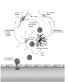
Brain mononuclear phagocytes (MP; perivascular macrophages and microglia) in nervous system health and disease. (A, top panel) Under steady state conditions, microglia secrete neurotrophic factors and engage other glial elements to promote tissue homeostasis. (B, bottom panel) During disease states (for example, Parkinson's and Alzheimer's disease), MP inflammatory responses damage the BBB, increase oxidative stress and release pro-inflammatory and pro-apoptotic cytokines and other neurotoxic factors that affect neuronal damage or dropout. The damage and stress signals enhance microglial activation, resulting in positive feedback in the release of chemokines and cytotoxic cytokines that cause further ingress of immune cells into the brain and expand inflammatory responses.
Figure 2
Figure 2
Neuroinflammatory and oxidative stress pathways in PD pathogenesis. Free radicals can arise several diverse ways, such as glial cell activation, mitochondrial dysfunction and protein aggregation. Microglial derived NO and superoxide species react in extracellular spaces to form peroxynitrite. Peroxynitrite readily crosses cell membranes where it contributes to lipid peroxidation, DNA damage and nitrotyrosine formation in α-synuclein and other cellular proteins. Damaged proteins are targeted to cellular proteosomes for degradation via the ubiquitination pathway. Excess NO produced by activated microglia can lead to S-nitrosylation of cellular proteins, including parkin. Such modifications may diminish E3 ubiquitin ligase activity necessary for efficient protein turnover by proteosomes. Excessive protein damage caused by oxidants and disruptions in the ubiquitin pathways may overload or inhibit protein degradation quality control measures leading to the accumulation of damaged proteins in cells. When reactive species exceed anti-oxidant defenses, oxidative stress is generated; destroying molecular structures, such as proteins, lipids and DNA, causing irreversible and detrimental damage, neuronal cell injury and death. Adapted from Gao et al. [261].
Figure 3
Figure 3
Diffusion tensor imaging of brain in a mouse model of PD. (A) Color encoding of the direction of the primary eigenvalue of the diffusion tensor is used to identify anatomical regions for analysis. (B) Results of quantitation of apparent diffusion coefficient of water (ADCω) and fractional anisotropy (FA) in striatum, corpus callosum, and substantia nigra in mouse brain before and after treatment with MPTP. No significant differences were found in the striatum and corpus callosum. Substantia nigra from mice treated with MPTP exhibited a significant increase in ADCω (P = 0.036) and decrease in FA (P = 0.002).
Figure 4
Figure 4
Cop-1 induced T cell-mediated neuroprotection in a PD model. In MPTP-intoxicated mice, regulatory T cells infiltrate the inflamed nigrostriatal pathway where they encounter cross-reactive self-antigens (myelin basic protein) presented in the context of MHC by resident microglial cells. Activated T cells secrete anti-inflammatory cytokines such as IL-4, IL-10, and TGF-β that suppress toxic microglial activities. Neurotrophin expression may occur directly from T cells or T cell derived IL-4 and IL-10 may induce neurotrophin production in neighboring glia. These activities lead to neuroprotection indirectly by suppression of microglial responses and directly through the local delivery of neurotrophins.

Similar articles

Cited by

References

    1. Mayeux R. Epidemiology of neurodegeneration. Annu Rev Neurosci. 2003;26:81–104. - PubMed
    1. Fahn S, Przedborski S. Parkinsonism. In: Rowland LP, editor. Merritt's Neurology. Lippincott Williams & Wilkins; New York: 2000. pp. 679–693.
    1. Fahn S, Sulzer D. Neurodegeneration and neuroprotection in Parkinson disease. NeuroRx. 2004;1:139–54. - PMC - PubMed
    1. Hornykiewicz O, Kish SJ. Biochemical pathophysiology of Parkinson's disease. Adv Neurol. 1987;45:19–34. - PubMed
    1. Clarke G, Collins RA, Leavitt BR, Andrews DF, Hayden MR, Lumsden CJ, et al. A onehit model of cell death in inherited neuronal degenerations. Nature. 2000;406:195–9. - PubMed

LinkOut - more resources