. 2008 Jan;118(1):294-305.
doi: 10.1172/JCI32103.

Adoptive transfer of effector CD8+ T cells derived from central memory cells establishes persistent T cell memory in primates

Affiliations

Adoptive transfer of effector CD8+ T cells derived from central memory cells establishes persistent T cell memory in primates

Carolina Berger et al. J Clin Invest. 2008 Jan.

Abstract

The adoptive transfer of antigen-specific T cells that have been expanded ex vivo is being actively pursued to treat infections and malignancy in humans. The T cell populations that are available for adoptive immunotherapy include both effector memory and central memory cells, and these differ in phenotype, function, and homing. The efficacy of adoptive immunotherapy requires that transferred T cells persist in vivo, but identifying T cells that can reproducibly survive in vivo after they have been numerically expanded by in vitro culture has proven difficult. Here we show that in macaques, antigen-specific CD8(+) T cell clones derived from central memory T cells, but not effector memory T cells, persisted long-term in vivo, reacquired phenotypic and functional properties of memory T cells, and occupied memory T cell niches. These results demonstrate that clonally derived CD8+ T cells isolated from central memory T cells are distinct from those derived from effector memory T cells and retain an intrinsic capacity that enables them to survive after adoptive transfer and revert to the memory cell pool. These results could have significant implications for the selection of T cells to expand or to engineer for adoptive immunotherapy of human infections or malignancy.


Figures

Figure 1
Figure 1. Isolation and gene marking of TCM- and TEM-derived CMV-specific CD8+ T cell clones for adoptive transfer.
(A) CMV IE-specific T cells are present in TCM and TEM subsets of CD8+ peripheral blood lymphocytes. PBMCs from macaque 02258 were stained with mAbs to CD28, Fas, and CD8 to identify CD28+CD8+Faslo TN, CD28+CD8+Fashi TCM, and CD28CD8+Fashi TEM fractions (left panel) and assayed by cytokine flow cytometry after stimulation with a CMV peptide (right panels). (B) Sorted CD62L+CD28+CD8+Faslo TN (top panel), CD62L+CD28+CD8+ Fashi TCM (middle panel), and CD62LCD28CD8+Fashi TEM (lower panel) T cells were stimulated with autologous CMV IE peptide-pulsed monocytes and assayed for cytolytic activity against peptide-pulsed (filled triangles) or unpulsed target cells (open circles). (C) Isolation of TCM- and TEM-derived CMV-specific CD8+ T cell clones. CD8+CD62L and CD8+CD62L+ T cell subsets were cultured with peptide-pulsed monocytes. At day 7, T cell clones were isolated by limiting dilution, transduced to express ΔCD19 or CD20, and expanded for adoptive transfer. (D) Design of the retroviral vector constructs. MPSV-LTR, myeloproliferative sarcoma virus retroviral long terminal repeat; ψ+, extended packaging signal; PRE, woodchuck hepatitis virus posttranscriptional regulatory element; ΔCD19, truncated macaque CD19 cDNA; CD20, full-length macaque CD20 cDNA. (E) Selection of ΔCD19- and CD20-modified CD8+ T cell clones. Transduced T cells were enriched for ΔCD19 or CD20 expression using immunomagnetic beads and analyzed by flow cytometry to assess purity (right panels). Unmodified T cells (left panels) served as control. The percentages of CD8+ T cells positive for CD19 or CD20 are indicated.
Figure 2
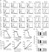
Figure 2. Phenotypic and functional characterization of TCM- and TEM-derived CMV-specific CD8+ T cell clones.
(A) Expression of CD62L, CCR7, CD28, CD127, granzyme B, and perforin (bold line) on individual TCM- and TEM-derived clones from 2 macaques. The dotted line indicates results from staining with an isotype control. Inset values represent the MFI. The data are shown for pairs of TE clones used for adoptive transfer in macaques 02269 and 02258 and is representative of data for all clones used for adoptive transfer. (B) Cytotoxic activity of each pair of TEM-derived (filled triangles) and TCM-derived (filled squares) clones was examined at an effector-to-target ratio (E/T ratio) of 20:1 using autologous peptide-pulsed target cells. The peptide sequences were KKGDIKDRV (macaque 02269), ATTRSLEYK (macaque 02258), and EEHVKLFFK (macaque A99171). (C) In vitro growth of CMV-specific CD8+ T cell clones. TEM-derived (filled triangles) and TCM-derived (filled squares) clones used for adoptive transfer were stimulated with anti-CD3 and anti-CD28 mAbs, γ-irradiated feeder cells, and IL-2 (50 U/ml), and cell growth was measured by counting viable cells. (D) Telomere length in TCM- and TEM-derived CMV-specific T cell clones. The median telomere length of duplicate samples was measured by automated flow-FISH in peripheral blood T lymphocytes, in the infused T cell clones (iTEM and iTCM), and in each of 2 additional randomly selected TEM- and TCM-derived T cell clones from each macaque.
Figure 3
Figure 3. Persistence and migration of TEM- and TCM-derived CD8+ TE clones in PBMCs, BM, and LNs following adoptive transfer.
(A and B) ΔCD19-modified TEM-derived (A) and TCM-derived (B) clones were transferred to macaque 02269 in separate infusions at a cell dose of 3 × 10⁸/kg, and samples of PBMCs, BM, and LNs were collected before (pre) and at the indicated times following infusion. The frequency of transferred CD19+ T cells was determined by flow cytometry and by PCR for vector sequences. Left: Percentage of CD19+CD8+ T cells by flow cytometry in PBMCs, BM, and LNs before and after infusion of the TEM-derived (A) and TCM-derived (B) clones. Cells were gated on CD3+CD8+ T cells. Right: Absolute numbers of CD19+CD8+ T cells/μl of blood determined by flow cytometry (gray bars; left y axis) and vector-positive T cells/106 PBMCs (filled diamonds; right y axis). (C and D) ΔCD19-modified TCM-derived and CD20-modified TEM-derived clones were transferred to macaque 02258 in separate infusions at a cell dose of 6 × 10⁸/kg. Aliquots of blood, BM, and LNs obtained at the indicated times were analyzed by flow cytometry and PCR to detect transferred CD19+CD8+ (C) or CD20+CD8+ (D) cells, respectively. Left: Percentage of CD8+ T cells that expressed ΔCD19 (C) or CD20 (D) in PBMCs, BM, and LNs. Cells were gated on CD3+CD8+ T cells. Right: Absolute number of marked CD8+ T cells/μl of blood (gray bars; left y axis) and vector-positive T cells/106 PBMCs (filled diamonds; right y axis).
Figure 4
Figure 4. CD8+ TCM-derived clones exhibit improved survival in IL-15 in vitro and decreased apoptosis in vivo.
(A) Aliquots of TEM- and TCM-derived clones used for adoptive transfer were plated at the end of a 14-day stimulation cycle in medium alone (open triangles), IL-2 (1 ng/ml) (filled diamonds), or IL-15 (1 ng/ml) (filled squares), and viability was determined using Trypan blue dye exclusion. Data are representative of 13 TCM- and 11 TEM-derived clones from 3 macaques. (B) Expression of IL-15Rα, IL-2Rβ, and IL-2Rγ on TCM-derived (bold lines) and TEM-derived (black lines) clones transferred to macaque 02269 was measured by flow cytometry on days 13–14 after stimulation (dotted lines, isotype control mAb). Data are representative of clones administered to macaques 02258 and A99171. (C) Bcl-xl and Bcl-2 expression on 3 TEM-derived (white bars) and TCM-derived (gray bars) clones transferred to macaques 02269, 02258, and A99171 analyzed 14 days after stimulation. Mean ± SD of the MFI is shown on the y axis. (D) Apoptosis of TEM- and TCM-derived clones in vivo. The percent of CD19+CD8+ T cells in PBMCs that were Annexin V+ and/or PI+ 1 day after infusion of ΔCD19+ TEM-derived or TCM-derived T cell clones to macaque A99171 was determined directly and after 24-hour culture. Samples were gated on CD8+CD19 (white bars) and CD8+CD19+ T cells (black bars). Hatched bars show the T cells used for infusion. (E) Persistence and migration of TEM-derived and TCM-derived TE clones in macaque A99171. PBMC, BM, and LN cells obtained at indicated times were analyzed by flow cytometry to detect transferred CD19+CD8+ TE in samples gated on CD3+CD8+.
Figure 5
Figure 5. Persistence and migration of a TEM-derived CD8+ TE clone in PBMCs and tissue sites following adoptive transfer.
(A) Persistence and migration of a ΔLNGFR-modified TEM-derived T cell clone in PBMCs. The ΔLNGFR+ T cell clone was transferred at a cell dose of 6 × 10⁸/kg. Aliquots of blood were collected at the indicated times and analyzed by PCR for the frequency of transferred ΔLNGFR+ T cells. Shown are the absolute numbers of vector-positive T cells/106 PBMCs (gray bars) before and at intervals after infusion. (B) A necropsy was performed on day 10 after the T cell infusion. Samples of DNA were isolated from various tissues as indicated and examined for the presence of the transferred vector-positive cells (black bars) using a real-time PCR assay. DNA isolated from ΔLNGFR+ T cells was serially diluted 1:10 in water prior to amplification with the appropriate primer set. The real-time PCR assay to detect β-actin (gray bars) was used as a control to detect genomic DNA. The threshold cycle is shown for each sample.
Figure 6
Figure 6. Adoptively transferred TCM-derived TE reexpress markers of TCM in vivo and persist in memory cell niches.
(A) Expression of CD62L on TEM-derived (upper panels) or TCM-derived (lower panels) TE clones after adoptive transfer. Samples of PBMCs were obtained before and on day 3 after the infusion of TCM- and TEM-derived clones and analyzed by flow cytometry after gating on CD3+CD8+ T cells. The percentage of T cells that expressed the ΔCD19 or CD20 marker gene and CD62L is shown in the upper right quadrant of each panel. (BE) A major fraction of CD8+ T cells that persist after adoptive transfer acquire phenotypic markers of TCM and reside in LNs. Aliquots of PBMCs, LNs, and BM were obtained from macaque 02258 at days 14 and 56 after infusion of the ΔCD19-modified TCM-derived clone. The expression of phenotypic markers of TCM, including CD62L (B), CCR7 (C), CD28 (D), and CD127 (E) on the subset of transferred CD19+ T cells was determined by flow cytometry after gating on CD3+CD8+ cells.
Figure 7
Figure 7. Adoptively transferred CD8+ T cells exhibit functional properties of TM.
(A) IFN-γ production. PBMCs obtained from macaque 02269 before and 14 days following infusion of a ΔCD19+ CMV-specific T cell clone were stimulated with medium, PMA/ionomycin, or peptide antigen and examined by cytokine flow cytometry. Data are gated on CD3+CD8+ cells and are representative of results from macaque 02258. (B) Transferred T cells that reexpress CD62L lack direct cytotoxicity but acquire cytotoxic function after stimulation. Left: PBMCs obtained 14–70 days after infusion of a TCM-derived CD19+CD8+ clone to macaque 02258 were pooled, sorted into CD19+CD62LCD8+ and CD19+CD62L+CD8+ fractions (purity >80%), and examined for lysis of autologous unpulsed (white bars) or peptide-pulsed target cells (black bars) (E/T ratio, 5:1). The cultured TCM-derived ΔCD19+CD8+ clone served as positive control for lysis. Right: Sorted CD19+CD62LCD8+ and CD19+CD62L+CD8+ T cells were stimulated using anti-CD3 and anti-CD28 mAbs for 14 days and then assayed for lysis of peptide-pulsed target cells (E/T ratio, 5:1). (C) Granzyme B expression. PBMCs and the transferred TCM-derived clone were stained with mAbs to CD62L, CD8, and CD19 as well as intracellular granzyme B. Cells were analyzed by flow cytometry after gating on CD62L+CD8+, CD62LCD8+, CD19+CD62L+CD8+, and CD19+CD62LCD8+ cells. (D) Proliferation. PBMCs obtained from macaque 02269 on days 14–70 after infusion were sorted into CD19+CD62L+CD8+ (left panel) and CD19+CD62LCD8+ subsets (right panel), labeled with CFSE, and stimulated with peptide-pulsed APCs as described in Methods. After 5 days, CFSE dilution was assessed by flow cytometry after gating on CD19+CD3+CD8+ cells. M3 gate, proportion of cells that have undergone more than 5 divisions.
Figure 8
Figure 8. Adoptively transferred T cells expand in vivo to antigen stimulation.
(A) T-APCs pulsed with IE peptide are lysed by IE-specific CD8+ T cells. T-APCs generated from macaque A99171 were pulsed with peptide (filled squares) or media alone (open squares), labeled with 51Chromium, and used as targets for the autologous IE-specific CD8+ T cell clone that was used for adoptive transfer. (B) Expansion of endogenous and transferred CMV-specific T cells by infusion of T-APCs. A dose of 1 × 10⁷ T-APCs/kg was administered to macaque A99171 nine weeks after infusion of a CD19+ TCM-derived clone. The number of CD3+CD8+ T cells and CD19+CD8+ T cells/μl of blood was measured by flow cytometry prior to and on indicated days following the T-APC administration. The number of endogenous IE-specific CD8+ T cells was measured by cytokine flow cytometry at the same time points after gating on CD19CD3+CD8+ cells. The data show the fold increase in the absolute numbers of CD3+CD8+ T cells (white bars), CD19+CD8+ T cells (gray bars), and CD19 IE-specific CD8+ T cells (black bars) after the T-APC infusion.

Similar articles

Cited by

References

    1. Cheever M.A., Greenberg P.D., Fefer A. Specificity of adoptive chemoimmunotherapy of established syngeneic tumors. J. Immunol. 1980;125:711–714. - PubMed
    1. Pahl-Seibert M.-F., et al. Highly protective in vivo function of cytomegalovirus IE1 epitope-specific memory CD8 T cells purified by T-cell receptor-based cell sorting. J. Virol. 2005;79:5400–5413. - PMC - PubMed
    1. Riddell S.R., et al. Restoration of viral immunity in immunodeficient humans by the adoptive transfer of T cell clones. Science. 1992;257:238–241. - PubMed
    1. Walter E.A., et al. Reconstitution of cellular immunity against cytomegalovirus in recipients of allogeneic bone marrow by transfer of T-cell clones from the donor. N. Engl. J. Med. 1995;333:1038–1044. - PubMed
    1. Rooney C.M., et al. Infusion of cytotoxic T cells for the prevention and treatment of Epstein-Barr virus-induced lymphoma in allogeneic transplant recipients. Blood. 1998;92:1549–1555. - PubMed

Publication types