Oxygen reperfusion damage in an insect
- PMID: 18060061
- PMCID: PMC2092388
- DOI: 10.1371/journal.pone.0001267
Oxygen reperfusion damage in an insect
Abstract
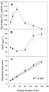
The deleterious effects of anoxia followed by reperfusion with oxygen in higher animals including mammals are well known. A convenient and genetically well characterized small-animal model that exhibits reproducible, quantifiable oxygen reperfusion damage is currently lacking. Here we describe the dynamics of whole-organism metabolic recovery from anoxia in an insect, Drosophila melanogaster, and report that damage caused by oxygen reperfusion can be quantified in a novel but straightforward way. We monitored CO(2) emission (an index of mitochondrial activity) and water vapor output (an index of neuromuscular control of the spiracles, which are valves between the outside air and the insect's tracheal system) during entry into, and recovery from, rapid-onset anoxia exposure with durations ranging from 7.5 to 120 minutes. Anoxia caused a brief peak of CO(2) output followed by knock-out. Mitochondrial respiration ceased and the spiracle constrictor muscles relaxed, but then re-contracted, presumably powered by anaerobic processes. Reperfusion to sustained normoxia caused a bimodal re-activation of mitochondrial respiration, and in the case of the spiracle constrictor muscles, slow inactivation followed by re-activation. After long anoxia durations, both the bimodality of mitochondrial reactivation and the recovery of spiracular control were impaired. Repeated reperfusion followed by episodes of anoxia depressed mitochondrial respiratory flux rates and damaged the integrity of the spiracular control system in a dose-dependent fashion. This is the first time that physiological evidence of oxygen reperfusion damage has been described in an insect or any invertebrate. We suggest that some of the traditional approaches of insect respiratory biology, such as quantifying respiratory water loss, may facilitate using D. melanogaster as a convenient, well-characterized experimental model for studying the underlying biology and mechanisms of ischemia and reperfusion damage and its possible mitigation.
Conflict of interest statement
Figures


Similar articles
-
Effects of temperature on responses to anoxia and oxygen reperfusion in Drosophila melanogaster.J Exp Biol. 2011 Apr 15;214(Pt 8):1271-5. doi: 10.1242/jeb.052357. J Exp Biol. 2011. PMID: 21430203
-
Intricate but tight coupling of spiracular activity and abdominal ventilation during locust discontinuous gas exchange cycles.J Exp Biol. 2018 Mar 15;221(Pt 6):jeb174722. doi: 10.1242/jeb.174722. J Exp Biol. 2018. PMID: 29386224
-
Spiracular fluttering decouples oxygen uptake and water loss: a stochastic PDE model of respiratory water loss in insects.J Math Biol. 2022 Apr 24;84(6):40. doi: 10.1007/s00285-022-01740-4. J Math Biol. 2022. PMID: 35461398
-
Control of the respiratory pattern in insects.Adv Exp Med Biol. 2007;618:211-20. doi: 10.1007/978-0-387-75434-5_16. Adv Exp Med Biol. 2007. PMID: 18269199 Review.
-
The respiratory basis of locomotion in Drosophila.J Insect Physiol. 2010 May;56(5):543-50. doi: 10.1016/j.jinsphys.2009.04.019. Epub 2009 Jun 2. J Insect Physiol. 2010. PMID: 19446563 Review.
Cited by
-
Hormetic benefits of prior anoxia exposure in buffering anoxia stress in a soil-pupating insect.J Exp Biol. 2018 Mar 19;221(Pt 6):jeb167825. doi: 10.1242/jeb.167825. J Exp Biol. 2018. PMID: 29367272 Free PMC article.
-
Interactions between Controlled Atmospheres and Low Temperature Tolerance: A Review of Biochemical Mechanisms.Front Physiol. 2011 Dec 2;2:92. doi: 10.3389/fphys.2011.00092. eCollection 2011. Front Physiol. 2011. PMID: 22144965 Free PMC article.
-
Subtropical hibernation in juvenile tegu lizards (Salvator merianae): insights from intestine redox dynamics.Sci Rep. 2018 Jun 19;8(1):9368. doi: 10.1038/s41598-018-27263-x. Sci Rep. 2018. PMID: 29921981 Free PMC article.
-
Anisotropic shrinkage of insect air sacs revealed in vivo by X-ray microtomography.Sci Rep. 2016 Sep 1;6:32380. doi: 10.1038/srep32380. Sci Rep. 2016. PMID: 27580585 Free PMC article.
-
An Efficient and Reliable Assay for Investigating the Effects of Hypoxia/Anoxia on Drosophila.Neurosci Bull. 2018 Apr;34(2):397-402. doi: 10.1007/s12264-017-0173-7. Epub 2017 Sep 2. Neurosci Bull. 2018. PMID: 28866769 Free PMC article.
References
-
- Idris AH, Roberts J, Caruso L, Showstark M, Layon J, et al. Oxidant injury occurs rapidly after cardiac arrest, cardiopulmonary resuscitation, and reperfusion. Crit Care Med. 2005;33:2043–2048. - PubMed
-
- Hermes-Lima M, Zenteno-Savin T. Animal response to drastic changes in oxygen availability and physiological oxidative stress. Comp Biochem Physiol C Comp Pharmacol. 2002;133:537–556. - PubMed
-
- Storey KB. 2004. Functional Metabolism: Regulation and Adaptation: Wiley-IEEE. p. 616.
-
- Joanisse DR, Storey KB. Oxidative stress and antioxidants in overwintering larvae of cold-hardy goldenrod gall insects. J Exp Biol. 1996;199:1483–1491. - PubMed
Publication types
MeSH terms
Substances
LinkOut - more resources
Full Text Sources
Molecular Biology Databases

