. 2007 Dec 5;2(12):e1269.
doi: 10.1371/journal.pone.0001269.

The mutant form of lamin A that causes Hutchinson-Gilford progeria is a biomarker of cellular aging in human skin

Affiliations

The mutant form of lamin A that causes Hutchinson-Gilford progeria is a biomarker of cellular aging in human skin

Dayle McClintock et al. PLoS One. .

Abstract

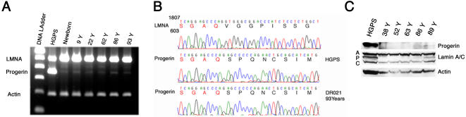
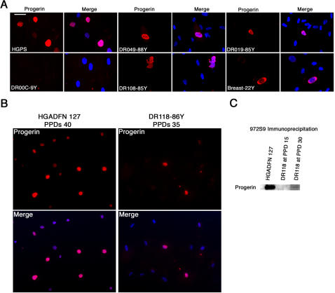
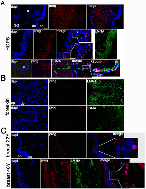
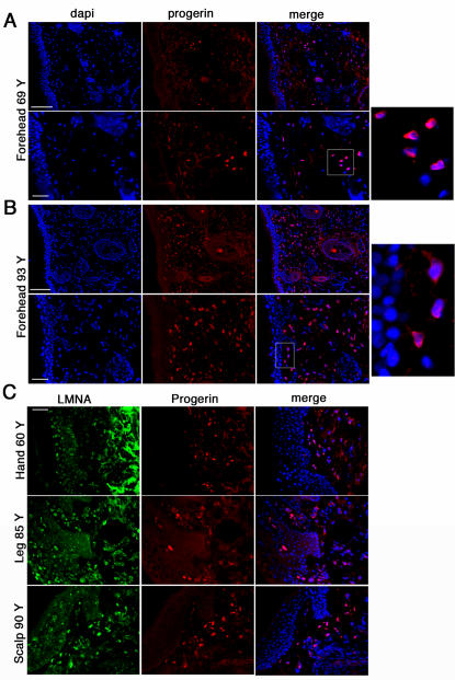
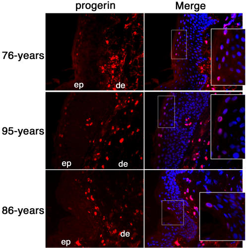
Hutchinson-Gilford progeria syndrome (HGPS, OMIM 176670) is a rare disorder characterized by accelerated aging and early death, frequently from stroke or coronary artery disease. 90% of HGPS cases carry the LMNA G608G (GGC>GGT) mutation within exon 11 of LMNA, activating a splice donor site that results in production of a dominant negative form of lamin A protein, denoted progerin. Screening 150 skin biopsies from unaffected individuals (newborn to 97 years) showed that a similar splicing event occurs in vivo at a low level in the skin at all ages. While progerin mRNA remains low, the protein accumulates in the skin with age in a subset of dermal fibroblasts and in a few terminally differentiated keratinocytes. Progerin-positive fibroblasts localize near the basement membrane and in the papillary dermis of young adult skin; however, their numbers increase and their distribution reaches the deep reticular dermis in elderly skin. Our findings demonstrate that progerin expression is a biomarker of normal cellular aging and may potentially be linked to terminal differentiation and senescence in elderly individuals.


Conflict of interest statement

Competing Interests: The authors have declared that no competing interests exist.

Figures

Figure 1
Figure 1. Progerin expression in human skin.
A, RT-PCR analysis of HGPS cells and human skin biopsies of indicated age, primers amplifying wild-type and progerin transcripts. B, Direct sequencing of a short portion within exon 11 of wild type lamin A (LMNA), and progerin transcripts from HGPS and 93-year-old subjects. C, Western blot analysis of protein extracted from skin biopsies of indicated age with anti-progerin 972S9, anti-lamin A/C and anti-actin antibodies.
Figure 2
Figure 2. Progerin expression in primary dermal fibroblast cultures.
A, Immunofluorescence microscopy on primary dermal fibroblasts from an HGPS subject and unaffected individuals of indicated ages with rabbit monoclonal anti-progerin antibody. B, Immunofluoresence detection of progerin in HGADFN 127 (HGPS individual) and DR118 (86-year-old individual) at late PPDs. C, Western blot analysis of nuclear protein extracts derived from fibroblast cultures HGADFN 127 (HGPS) at PPD 20 and DR118 (86-year-old female) at PPD 15 and 30, respectively, were immunoprecipitated with anti-progerin mAb 972S9 and the corresponding Western blot was probed with the same antibody.
Figure 3
Figure 3. In situ localization of progerin on human skin sections derived from a subject with HGPS and from unaffected individuals.
A, HGPS skin sections immunostained with anti-progerin (prog), anti-lamin A (LMNA) antibody, or anti-α smooth muscle actin antibody (αSMA) and counterstained with a DNA stain (dapi). Morphologic entities are indicated: epidermis (ep), dermis (de), sweat glands (SG), capillary (c), arrector pili muscle (a), and hair follicle (h). B, Newborn foreskin sections immunolabelled with anti-progerin or anti-αSMA antibody. C, Breast skin sections from 22- and 46-year-old female subjects probed with anti-progerin and lamin A. The respective double or triple merged signals are indicated.
Figure 4
Figure 4. Progerin accumulation in human elderly skin biopsy sections.
A, Forehead skin section from a 69-year-old individual probed with anti-progerin antibody and counterstained with dapi. Bars correspond to 100 and 50 µm, respectively. B, Forehead skin section from a 93-year-old donor. C, Skin sections from different body sites as indicated were probed with anti-progerin and anti- lamin A (LMNA) antibodies. Bar, 50 µm. Merged images are indicated.
Figure 5
Figure 5. Progerin detection in a subset of terminally differentiated keratinocytes.
Left panels correspond to anti-progerin monoclonal antibody staining of skin sections derived from individuals of indicated age. Right panels correspond to the merged signal of dapi and progerin signals. Square indicates the zoomed in region of the epidermis. ep denotes epidermis, and de the dermis.

References

    1. Aebi U, Cohn J, Buhle L, Gerace L. The nuclear lamina is a meshwork of intermediate-type filaments. Nature (London) 1986;323:560–564. - PubMed
    1. Hutchinson CJ, Worman HJ. A-type lamins: guardians of the soma? Nature Cell Biology. 2004;6:1062–1067. - PubMed
    1. Lin F, Worman HJ. Structural organization of the human gene (LMNB1) encoding nuclear lamin B1. Genomics. 1995;27:230–236. - PubMed
    1. Wydner KL, McNeil JA, Lin F, Worman HJ, Lawrence JB. Chromosomal assignment of human nuclear envelope protein genes LMNA, LMNB1, and LBR by fluorescence in situ hybridization. Genomics. 1996;32:474–478. - PubMed
    1. Furukawa K, Hotta Y. cDNA cloning of a germ cell specific lamin B3 from mouse spermatocytes and analysis of its function by ectopic expression in somatic cells. EMBO Journal. 1993;12:97–106. - PMC - PubMed

Publication types

MeSH terms