. 2007 Nov 28;1(2):e34.
doi: 10.1371/journal.pntd.0000034.

Optimization of topical therapy for Leishmania major localized cutaneous leishmaniasis using a reliable C57BL/6 Model

Affiliations

Optimization of topical therapy for Leishmania major localized cutaneous leishmaniasis using a reliable C57BL/6 Model

Hervé Lecoeur et al. PLoS Negl Trop Dis. .

Abstract

Background: Because topical therapy is easy and usually painless, it is an attractive first-line option for the treatment of localized cutaneous leishmaniasis (LCL). Promising ointments are in the final stages of development. One main objective was to help optimize the treatment modalities of human LCL with WR279396, a topical formulation of aminoglycosides that was recently proven to be efficient and safe for use in humans.

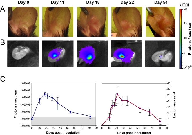
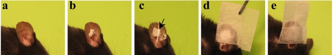
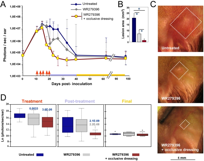
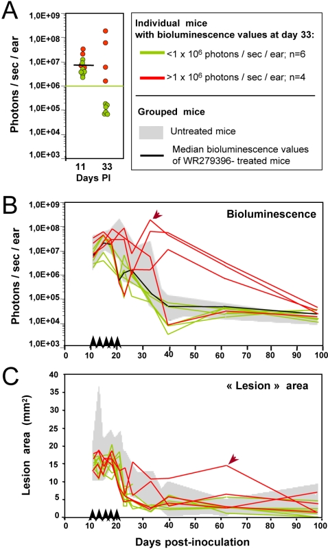
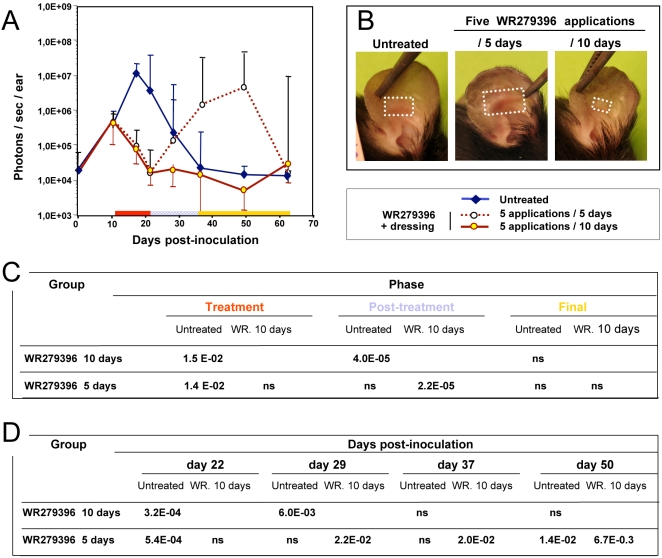
Methodology/principal findings: C57BL/6 mice were inoculated in the ear with luciferase transgenic L. major and then treated with WR279396. The treatment period spanned lesion onset, and the evolution of clinical signs and bioluminescent parasite loads could be followed for several months without killing the mice. As judged by clinical healing and a 1.5-3 log parasite load decrease in less than 2 weeks, the 94% efficacy of 10 daily applications of WR279396 in mice was very similar to what had been previously observed in clinical trials. When WR279396 was applied with an occlusive dressing, parasitological and clinical efficacy was significantly increased and no rebound of parasite load was observed. In addition, 5 applications under occlusion were more efficient when done every other day for 10 days than daily for 5 days, showing that length of therapy is a more important determinant of treatment efficacy than the total dose topically applied.

Conclusions/significance: Occlusion has a significant adjuvant effect on aminoglycoside ointment therapy of experimental cutaneaous leishmaniasis (CL), a concept that might apply to other antileishmanial or antimicrobial ointments. Generated in a laboratory mouse-based model that closely mimics the course of LCL in humans, our results support a schedule based on discontinuous applications for a few weeks rather than several daily applications for a few days.


Conflict of interest statement

HL, SG, GG, GMi and TL declare that no competing interests exist. PB and GMo were co-investigators in a Phase 2 clinical trial of WR279396 conducted under the co-sponsorship of the Office of The Surgeon General, Department of the Army, FDA IND number 50,098, HSRRB Protocol number A-97-68.1, and the Institut Pasteur, Paris, France. Their institution (Institut Pasteur) was awarded a grant from US Army Medical Research and Materiel Command (MRMC) for the execution of this clinical trial. MRMC is responsible for the development of WR279396.

Figures

Figure 1
Figure 1. In vivo bioluminescence imaging of Leishmania in the C57BL/6 ear model.
Simultaneous follow up of parasite load in the ear dermis and of lesion onset, features and cure in mice inoculated with luciferase-transgenic Leishmania major. 104 luciferase-expressing NIH 173 metacyclic promastigotes were inoculated into the dermis of the right ear of C57BL/6 mice (day 0) and followed for more than 80 days. The bioluminescent signal is displayed as a pseudo-colour image representing light intensities over the body surface area. Red represents the most intense signal while blue corresponds to the weakest one. A, B: Individual follow up of a representative mouse left without any ointment application. Clinical features (A) were simultaneously monitored with parasite load fluctuations assessed by the bioluminescence of luciferase-expressing parasites (B). (C) Bioluminescence quantification of the parasite load (left panel; photons/sec/ear; grey area = background measurement) and “lesion” area (right panel; mm2) were followed for 7 mice left without any ointment and depicted as medians +/−sd. Note the detection of a bioluminescence signal before any significant clinically detectable features. Of note, the so called “lesion” area measured between day 11 and day 15 was still made up of inflammatory processes free of any leukocyte infiltrates.
Figure 2
Figure 2. C57BL/6 mice given an application of WR279396 formulation under an occlusive dressing.
104 luciferase-expressing L. major metacyclic promastigotes were inoculated into the dermis of the right ear of C57BL/6 mice (day 0). (a, b) From day eleven post-L. major inoculation, the topical ointment WR279396 was applied directly to parasite-loaded ears and (c) then covered with an adhesive polyurethane dressing (arrow). (d,e) Two independent leaflets of surgical tape were applied directly on the occlusive dressing. This surgical tape permitted maintenance of the dressing and kept the formulation in contact with the parasite-loaded site for two days.
Figure 3
Figure 3. Earlier and sustained control of both parasite load and lesion healing post topical applications of WR 279396.
104 luciferase-expressing L. major metacyclic promastigotes were inoculated into the dermis of the right ear of C57BL/6 mice (day 0) and mice were followed for 3 months. We compared parasite load and clinical feature outcome in mice left without any ointment (group 1, blue line) or receiving the WR279396 ointment (one application ( ↑ ) every two days, over ten days either without dressing (group 2, grey line) or with an occlusive dressing (group 3, brown line) at 11 day post-inoculation of promastigotes. Total photon emission from the L. major-loaded site (A, photons/sec/ear) is depicted as medians ±sd. Lesion area (B; mm2) and representative pictures of C57BL/6 L. major loaded-ears (C), at day 28 post-inoculation, are presented. Significant differences are indicated as follows: * for P<0.001. White squares delimit the “lesion” area of groups 1, 2 and 3 respectively. The three periods of observation (treatment, post-treatment and final) are represented by a colour code. (D) Comparison of bioluminescence (natural logarithm (ln) of photons/sec/ear) for each group of mice during the period of observation. The box plot for each group, assessed by a two-way ANOVA, graphs the percentile and median of parasite loads. The ends of the box define the 25th and 75th percentiles, with a line at the median and errors bars defining the 10th and 90th percentiles. The dots outside the ends of the whiskers are outliers. The p-values are displayed on the top of each box with the following colour code: -blue indicates that the group under study is compared to the blue group-grey indicates that the group under study is compared to the grey group.
Figure 4
Figure 4. Individual follow up of bioluminescence (parasite load) and “lesion” area in mice treated with WR279396 without any dressing.
104 luciferase-expressing L. major metacyclic promastigotes were inoculated into the dermis of the right ear of C57BL/6 mice (n = 10; day 0) treated with WR279396 (5 applications (↑) for 10 days) at day 11 post-inoculation. The parasite load (photons/sec/ear) in individual mice (A, B) and the area (mm2; C) of the lesion displayed by the same mice were followed for 3 months. (A, B, C) Green colour assesses the profile in mice that controlled their ear parasite load, i.e. exhibiting a bioluminescence value<1×10⁶ photons/sec/ear at day 33 (green points in panel A and green lines in panels B and C). In contrast, red colour corresponds to mouse ears that display a high bioluminescence value at day 33 (>1×10⁶ photons/sec/ear; red points in panel A and red lines in panels B and C). Note that lesion area values (C) did not assess any clinical failure except in 1 mouse (arrow). Values obtained for control mice are shown in the grey areas indicated in each graph.
Figure 5
Figure 5. Determination of the most suitable regimen for WR279396 application under an occlusive dressing.
Eleven days post-inoculation of 104 luciferase-transfected L. major parasites, three groups of 7 mice were constituted: In the control group, mouse ears were left without any ointment (blue plain line), and in the other group WR279396 was applied on the mouse ear under an occlusive dressing with different application frequencies; either 5 applications/10 days (brown plain line) or 5 applications/5 days (brown dotted line). Bioluminescence evaluation of parasite load was performed over the course of 64 days post-inoculation (A). Note that the rebound of the parasite load in the every day-application group can be associated to a more severe lesion as illustrated by a representative mouse at day 36 post-inoculation (B/5 days). White squares delimit the “lesion” area. (C, D) Parasites loads were analysed with ANOVA whose two factors were the treatment (WR279396 10 days or 5 days) and the period of observation. Pair wise comparisons using t-tests were realized for each combination of factors.

References

    1. Desjeux P. Worldwide increasing risk factors for leishmaniasis. Medical Microbiology&Immunology. 2001;190:77–79. - PubMed
    1. Alvar J, Yactayo S, Bern C. Leishmaniasis and poverty. Trends in Parasitology. 2006;22:552–557. - PubMed
    1. Buffet P, Morizot G. Cutaneous leishmaniasis in France: towards the end of injectable therapy? Bull Soc Pathol Exot. 2003;96:383–388. - PubMed
    1. Magill AJ. Cutaneous leishmaniasis in the returning traveler. Infect Dis Clin North Am. 2005;19:241–266. - PubMed
    1. Bryceson A. Therapy in man. In: Peters W, Killick-Kendrick R, editors. The leishmaniases. London: Academic Press; 1987. pp. 847–869.

Publication types

LinkOut - more resources