Effects of cathepsins B and L inhibition on postischemic protein alterations in the brain
- PMID: 18060871
- PMCID: PMC3878606
- DOI: 10.1016/j.bbrc.2007.11.104
Effects of cathepsins B and L inhibition on postischemic protein alterations in the brain
Abstract
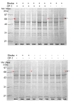
The effects of selective inhibition of cathepsins B and L on postischemic protein alterations in the brain were investigated in a rat model of middle cerebral artery occlusion (MCAO). Cathepsin B activity increased predominantly in the subcortical region of the ischemic hemisphere where the levels of collapsing mediator response protein 2, heat shock cognate 70 kDa protein, 60 kDa heat shock protein, protein disulfide isomerase A3 and albumin, were found to be significantly elevated. Postischemic treatment with Cbz-Phe-Ser(OBzl)-CHN(2), cysteine protease inhibitor 1 (CP-1), reduced infarct volume, neurological deficits and cathepsin B activity as well as the amount of heat shock proteins and albumin found in the brain. Our data strongly suggests that the decrease in heat shock protein levels and the significant reduction of serum albumin leakage into the brain following acute treatment with CP-1 is indicative of less secondary ischemic damage, which ultimately, is related to less cerebral tissue loss and improved neurological recovery of the animals.
Figures


References
-
- Lopez AD, Mathers CD, Ezzati M, Jamison DT, Murray CJ. Global and regional burden of disease and risk factors, 2001: systematic analysis of population health data. Lancet. 2006;367:1747–1757. - PubMed
-
- WHO. The World Health Report 2003. 2003. Online at http://www.who.int/whr/2003/en/Facts_and_Figures-en.pdf.
-
- Rosamond W, Flegal K, Friday G, et al. Heart disease and stroke statistics-2007 update. Circulation. 2007;115:e99–e110. - PubMed
-
- Siesjo B. Calcium and ischemic brain damage. Eur Neurol. 1986;25:45–56. - PubMed
-
- Ogata N, Yanekawa Y, Taki W, Kannagi R, Murachi T, Hamakubo T, Kikuchi H. Degradation of neurofilament protein in cerebral ischemia. J Neurosurg. 1989;70:103–107. - PubMed
Publication types
MeSH terms
Substances
Grants and funding
LinkOut - more resources
Full Text Sources
Research Materials
Miscellaneous

