Review
. 2008 Jan;44(1):31-43.
doi: 10.1016/j.yjmcc.2007.10.012. Epub 2007 Oct 25.

Mechanisms and potential therapeutic targets for ventricular arrhythmias associated with impaired cardiac calcium cycling

Affiliations
Review

Mechanisms and potential therapeutic targets for ventricular arrhythmias associated with impaired cardiac calcium cycling

Kenneth R Laurita et al. J Mol Cell Cardiol. 2008 Jan.

Abstract

The close relationship between life-threatening ventricular arrhythmias and contractile dysfunction in the heart implicates intracellular calcium cycling as an important underlying mechanism of arrhythmogenesis. Despite this close association, however, the mechanisms of arrhythmogenesis attributable to impaired calcium cycling are not fully appreciated or understood. In this report we review some of the current thinking regarding arrhythmia mechanisms associated with either abnormal impulse initiation (i.e. arrhythmia triggers) or impulse propagation (i.e. arrhythmia substrates). In all cases, the mechanisms are primarily related to dysfunction of calcium regulatory proteins associated with the sarcomere. These findings highlight the broad scope of arrhythmias associated with abnormal calcium cycling, and provide a basis for a causal relationship between cardiac electrical instability and contractile dysfunction. Moreover, calcium cycling proteins may provide much needed targets for novel antiarrhythmic therapies.


Figures

Figure 1
Figure 1
A simplified schematic of normal myocyte intracellular calcium cycling. Shown are the main proteins and their primary responsibility in the rapid increase of cytoplasm calcium (red) during systole and the subsequent decrease in cytoplasmic calcium levels (blue) during diastole. During steady state conditions, intracellular calcium homeostasis is maintained by an equal amount of calcium entering and exiting the cytoplasm for each beat. Shown below for reference is the time course of transmembrane potential and cytoplasmic calcium levels during a single beat.
Figure 2
Figure 2
Example of triggered activity and DADs in a Purkinje fiber during Digitalis administration. The arrow indicates the last paced beat, which is followed by two triggered (i.e. ectopic) beats and damped oscillations of membrane potential the represent DAD activity. Reproduced with permission.
Figure 3
Figure 3
Triggered activity in the canine wedge preparation under conditions of enhanced calcium entry. Shown in Panel A are representative action potentials (Vm) and calcium transients (Ca) recorded during baseline pacing (600ms) from the endocardium (left) and rapid pacing (180ms) followed by an abrupt halt in pacing from the same endocardial site (middle) and an epicardial site (right). After cessation of rapid pacing, an ectopic beat is evident (asterisk). In contrast to the epicardium, a delayed-afterdepolarization (DAD) and a spontaneous calcium release (SCR) occurred at the endocardium that did not elicit an ectopic beat. The activation map of the ectopic beat (Panel B) shows its origin (asterisk) close to the endocardium yet away from the site of stimulation (stimulus symbol). Overall experiments (Panel C), triggered activity (TA) occurred more frequently at the endocardium compared to other regions (p<0.001). Reproduced with permission.
Figure 4
Figure 4
Multiple simultaneous SCR events in the canine wedge preparation under conditions of enhanced calcium entry. In Panel A, a representative transmural map of SCR amplitude (SCRamp) reveals 2 “hot spots” of high SCR amplitude near the endocardium (a) and midmyocardial region (c). Panel B shows the ECG and calcium transients recorded at a, b, c and d. SCR event at site a had an earlier occurrence and larger amplitude compared to b and c. Note that no SCR was observed at d (epicardium). Reproduced with permission.
Figure 5
Figure 5
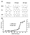
ECG, Vm, and CaF recordings at 3 different pacing rates (A). The number below each Vm and CaF recording represents APD (ms) and normalized CaF amplitude (%), respectively. The development of T-wave alternans was closely paralleled by APD-ALT and CaF-ALT. At 150 bpm (left), all signals were stable over time, and no alternans was observed. At 375 bpm (middle), subtle beat-to-beat alternans was observed, which was further increased at a pacing rate of 461 bpm. B, Representative experiment showing that the magnitude of APD-ALT and CaF-ALT increased as a function of pacing rate. Reproduced with permission.
Figure 6
Figure 6
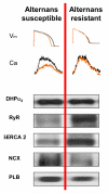
Molecular profile of alternans susceptible VS. resistant myocytes. Endocardial myocyte (left) undergoes significant Vm-ALT and Ca-ALT (black & orange traces) while under identical conditions the epicardial myocyte (right) does not. Western immunoblots show decreased RyR and SERCA2a in alternans susceptible myocytes. Reproduced with permission.
Figure 7
Figure 7
ECG and calcium transients near the epicardium (EPI) and endocardium (ENDO) recorded during an abrupt increase in pacing cycle length from 600 to 300 msec (arrow). Near ENDO and EPI, calcium transient alternans was persistent following 4 seconds of rapid pacing. At ENDO, where the decay of the calcium transient was slower (174 ms) compared to EPI (93 ms), the magnitude of calcium transient alternans was greater. At ENDO, the calcium transient amplitude for beat a was 68% larger than that for beat b. In contrast, at EPI the degree of calcium transient alternans was less obvious (9%). Reproduced with permission.

References

    1. Zheng ZJ, Croft JB, Giles WH, Mensah GA. Sudden cardiac death in the United States, 1989 to 1998. Circulation. 2001;104:2158–2163. - PubMed
    1. Solomon SD, Zelenkofske S, McMurray JJ, Finn PV, Velazquez E, Ertl G, et al. Sudden death in patients with myocardial infarction and left ventricular dysfunction, heart failure, or both. N Engl J Med. 2005;352:2581–8. - PubMed
    1. Bardy GH, Lee KL, Mark DB, Poole JE, Packer DL, Boineau R, et al. Amiodarone or an implantable cardioverter-defibrillator for congestive heart failure. N Engl J Med. 2005;352:225–237. - PubMed
    1. Tomaselli GF, Marbán E. Electrophysiological remodeling in hypertrophy and heart failure. Cardiovasc.Res. 1999;42:270–283. - PubMed
    1. Pruvot E, Katra RP, Rosenbaum DS, Laurita KR. Calcium cycling as a mechanism of repolarization alternans onset in the intact heart. Circulation. 2002;19:11–191. Abstract.