

Validation of a fiducial-based atlas localization method for deep brain stimulation contacts in the area of the subthalamic nucleus
- PMID: 18061273
- PMCID: PMC2267480
- DOI: 10.1016/j.jneumeth.2007.10.007
Validation of a fiducial-based atlas localization method for deep brain stimulation contacts in the area of the subthalamic nucleus
Abstract
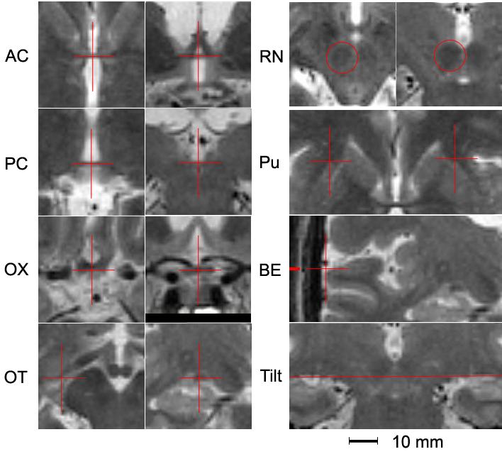
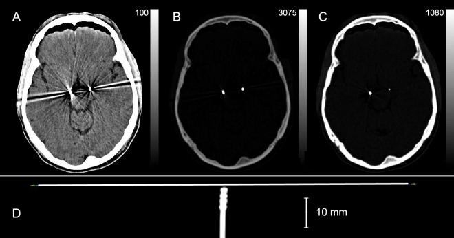
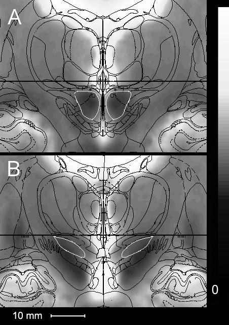
Differences in the location of active contacts with respect to the subthalamic nucleus (STN) may account for much variability in motor, psychiatric and cognitive responses to deep brain stimulation (DBS) in Parkinson disease (PD) patients. Because localization of STN based on hypointensity in T2-weighted MR images is unreliable and further limited by artifacts from the metal electrodes, we developed and validated a method to transform brain images into stereotactic space [Mai JK, Assheuer J, Paxinos G. Atlas of the Human Brain, 2nd ed. San Diego: Elsevier Academic; 2004] using reliably-identified anatomic fiducials identified in high-resolution T2-weighted pre-operative MR images. Average intraclass correlation between two raters for 29 PD patients was 0.93 for those fiducials used to define the atlas. Accuracy of the registration was tested by comparing the rater-identified centers of the red nuclei with their predicted locations from the fiducial-based atlas transformation. Mean discrepancies were 0.1, 0.9, and 0.0mm (x, y, z) with standard deviations of 0.9, 0.7 and 1.1mm, respectively. Because post-operative determination of contact location with respect to the STN is necessary due to possible shifting of electrodes during surgical placement, we identified active contacts on post-operative CT images and transformed their locations into stereotactic space. This method provides an accurate and reliable means for STN DBS contact localization.
Figures
References
-
- Bardinet E, Dormont D, Malandain G, Bhattacharjee M, Pidoux B, Saleh C, Cornu P, Ayache N, Agid Y, Yelnik J. Retrospective cross-evaluation of an histological and deformable 3D atlas of the basal ganglia on a series of Parkinsonian patients treated by deep brain stimulation. Medical Image Computing & Computer-Assisted Intervention: MICCAI. 2005;8:385–393. - PubMed
-
- Burn DJ, Troster AI. Neuropsychiatric complications of medical and surgical therapies for Parkinson’s disease. J Geriatr Psychiatry Neurol. 2004;17:172–180. - PubMed
-
- Castro FJS, Pollo C, Meuli R, Madeer P, Cuisenair O, Cuadra MB, Villemure J-G, Thiran J-P. A cross validation study of deep brain stimulation targeting: From experts to atlas-based, segmentatin-based and automatic registration algorithms. IEEE Trans Med Imag. 2006:1440–1450. - PubMed
-
- Christensen GE, Joshi SC, Miller MI. Volumetric transformation of brain anatomy. IEEE Trans Med Imag. 1997:864–877. - PubMed
-
- Deuschl G, Schade-Brittinger C, Krack P, Volkmann J, Schäfer H, Bötzel K, Daniels C, Deutschländer A, Dillmann U, Eisner W, Gruber D, Hamel W, Herzog J, Hilker R, Klebe S, Kloß M, Koy J, Krause M, Kupsch A, Lorenz D, Lorenzl S, Mehdorn HM, Moringlane JR, Oertel W, Pinsker MO, Reichmann H, Reuß A, Schneider G-H, Schnitzler A, Steude U, Sturm V, Timmermann L, Tronnier V, Trottenberg T, Wojtecki L, Wolf E, Poewe W, Voges J. A randomized trial of deep-brain stimulation for Parkinson’s disease. N Engl J Med. 2006;355:896–908. - PubMed
Publication types
MeSH terms
Grants and funding
LinkOut - more resources
Full Text Sources
