Review
. 2008 Jan;43(1):1-8.
doi: 10.1016/j.ceca.2007.10.006. Epub 2007 Dec 3.

Immuno-proteomic approach to excitation--contraction coupling in skeletal and cardiac muscle: molecular insights revealed by the mitsugumins

Affiliations
Review

Immuno-proteomic approach to excitation--contraction coupling in skeletal and cardiac muscle: molecular insights revealed by the mitsugumins

Noah Weisleder et al. Cell Calcium. 2008 Jan.

Abstract

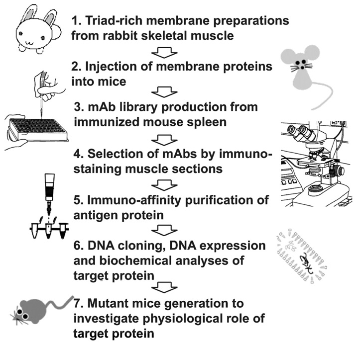
A comprehensive understanding of excitation-contraction (E-C) coupling in skeletal and cardiac muscle requires that all the major components of the Ca(2+) release machinery be resolved. We utilized a unique immuno-proteomic approach to generate a monoclonal antibody library that targets proteins localized to the skeletal muscle triad junction, which provides a structural context to allow efficient E-C coupling. Screening of this library has identified several mitsugumins (MG); proteins that can be localized to the triad junction in mammalian skeletal muscle. Many of these proteins, including MG29 and junctophilin, are important components in maintaining the structural integrity of the triad junction. Other triad proteins, such as calumin, play a more direct role in regulation of muscle Ca(2+) homeostasis. We have recently identified a family of trimeric intracellular cation-selective (TRIC) channels that allow for K(+) movement into the endoplasmic or sarcoplasmic reticulum to counter a portion of the transient negative charge produced by Ca(2+) release into the cytosol. Further study of TRIC channel function and other novel mitsugumins will increase our understanding of E-C coupling and Ca(2+) homoeostasis in muscle physiology and pathophysiology.


Figures

Fig. 1
Fig. 1
Using an immuno-proteomic monoclonal antibody library to discover novel proteins involved in E–C coupling and muscle physiology. This scheme illustrates the steps used to generate and screen a monoclonal antibody library against proteins that localize to the triad junction in skeletal muscle. Antibodies that show localization at the triad junction are used to isolated proteins for sequencing, which provides the sequence information to clone the genes of interest. Knockout mice can then be generated to study the physiological role of the gene of interest.
Fig. 2
Fig. 2
Molecular components of E–C coupling in striated muscle. While dihydropyridine receptor (DHPR) and ryanodine receptor (RyR) Ca2+ channels are major components of the E–C coupling machinery at the triad (in skeletal muscle) or dyad (in cardiac muscle) junctions, other accessory proteins are essential to maintain structural integrity and efficient RyR function. Mitsugumin-29 (MG29) acts in transverse (T)-tubule maintenance and can directly interact with RyR to modulate channel activity. Junctophilin (JP) maintains proper spacing between the T-tubule and sarcoplasmic reticulum (SR) by spanning the gap between the two membranes to physically link them together. Calumin may interact with Orai to regulate SOCE. Calsequestrin (CSQ) and sarcalumenin (SAR) are Ca2+ binding proteins that buffer SR Ca2+ stores. TRIC channels in the SR gate K+ movement to counteract transient negative potential produced by Ca2+ release from the SR. Other SR resident proteins, such as SERCA and K+ or Cl channels of unknown molecular identity, can also provide some aspects of this counter ion movement.
Fig. 3
Fig. 3
Various junctophilin mutations have been linked to human disease. Members of the JP family all contain a well-conserved series of MORN motifs (red) that interact with the PM and a transmembrane domain (TM, green) that inserts into the ER membrane. A divergent region that contains some alpha-helical sequences links these conserved domains. Mutations have been identified in JP-2 (arrows) that are linked to hypertrophic cardiomyopathy in different studies (Ref. [40] in black; Ref. [41] in blue). Numerous studies have identified triplet repeats in JP-3 exon 2a that result in Huntington’s disease-like symptoms. Sequences are based on human protein sequences for JP-1 (NP_065698), JP-2 (NP_065166), JP-3 (NP_065706) and JP-4 (NP_115828). Black numbers indicate the number of amino acid residues in each isoform. (For interpretation of the references to color in this figure legend, the reader is referred to the web version of the article.)
Fig. 4
Fig. 4
Calumin and STIM1 share a similar domain structure. Calumin contains a SP (signal peptide), an ER/SR lumen domain that can bind Ca2+ (lum), a single transmembrane domain (TM) with a long cytoplasmic (cyt) domain that contains a coiled coil region. Similarly, STIM1 contains a SP and Ca2+-binding EF-hand domain (EF) on a section of the protein localized within the lumen of the ER/SR, which also contains a sterile α-helix motif (SAM) domain. STIM1 also contains a single TM and a cytosolic coiled-coil domain. Black numbers indicate the number of amino acid residues in each isoform.

References

    1. Ma J, Pan Z. Junctional membrane structure and store operated calcium entry in muscle cells. Front Biosci. 2003;8:d242–d255. - PubMed
    1. Franzini-Armstrong C, Jorgensen AO. Structure and development of E–C coupling units in skeletal muscle. Annu. Rev. Physiol. 1994;56:509–534. - PubMed
    1. Meissner G. Ryanodine receptor/Ca2+ release channels and their regulation by endogenous effectors. Annu. Rev. Physiol. 1994;56:485–508. - PubMed
    1. Fill M, Copello JA. Ryanodine receptor calcium release channels. Physiol. Rev. 2002;82:893–922. - PubMed
    1. Farrell EF, Antaramian A, Benkusky N, Zhu X, Rueda A, Gomez AM, Valdivia HH. Regulation of cardiac excitation–contraction coupling by sorcin, a novel modulator of ryanodine receptors. Biol. Res. 2004;37:609–612. - PubMed