. 2008 Feb;151(2):235-43.
doi: 10.1111/j.1365-2249.2007.03555.x. Epub 2007 Dec 6.

Immunoregulatory factors in multiple sclerosis patients during and after pregnancy: relevance of natural killer cells

Affiliations

Immunoregulatory factors in multiple sclerosis patients during and after pregnancy: relevance of natural killer cells

L Airas et al. Clin Exp Immunol. 2008 Feb.

Abstract

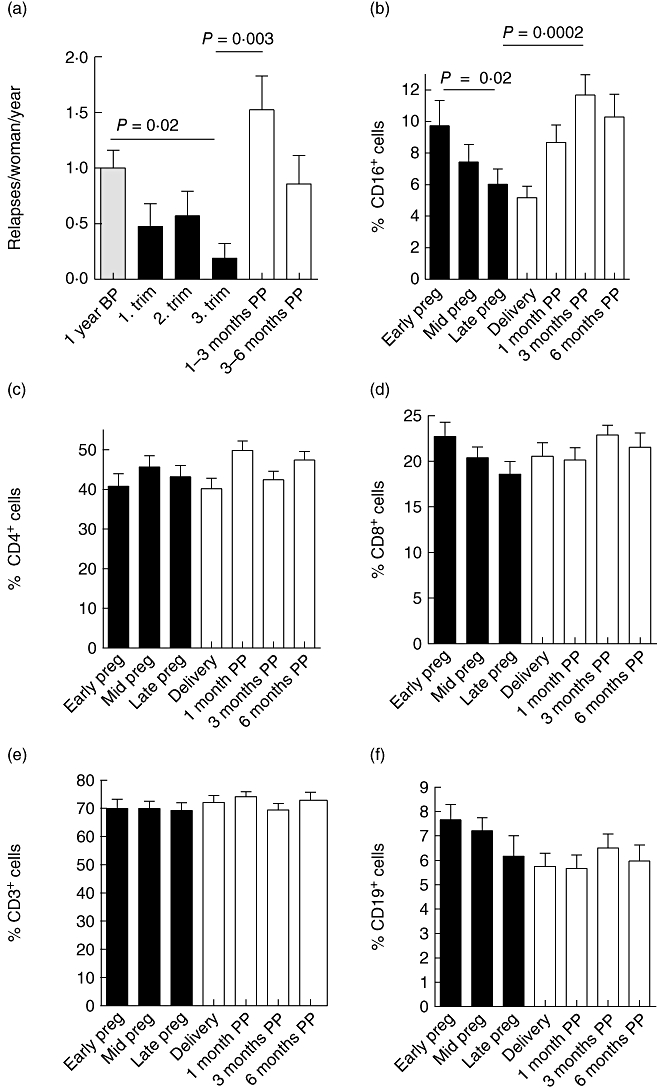
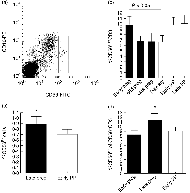
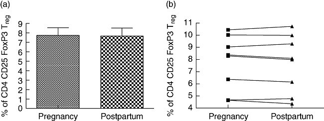
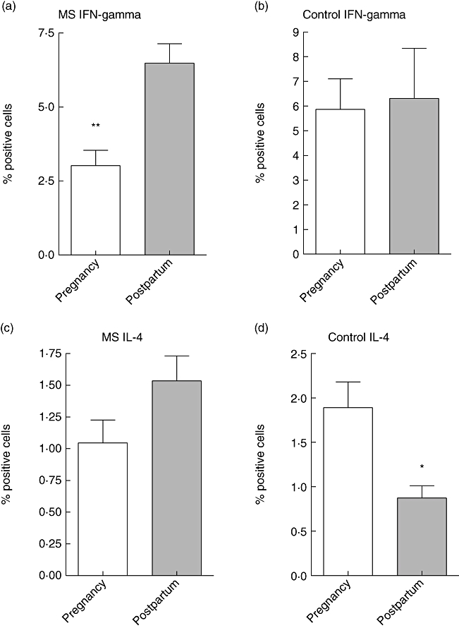
Multiple sclerosis (MS) ameliorates typically during pregnancy but after the delivery the relapse rate often increases. Our study was conducted to understand the immunoregulatory mechanisms accompanying this phenomenon. MS patients were followed-up prospectively during pregnancy and 6 months postpartum, with immunological characterization of the peripheral blood. Groups of age- and parity-matched healthy pregnant women, and age- and sex-matched non-pregnant women and non-pregnant MS patients were studied as controls. In our patient cohort, the annualized relapse rate was 1.0 +/- 1.0 relapses/woman/year (mean +/- standard deviation) during the year before pregnancy, but dropped to 0.2 +/- 0.9 during the third trimester (P = 0.02). After the delivery the relapse rate increased again to 1.4 +/- 1.9 (1-3 months postpartum versus third trimester P = 0.003). While percentages of peripheral blood CD3, CD4, CD8 and CD19 immune cell subsets were unchanged over the observation period, reduced disease activity during the last trimester was associated with a significant increase in the percentage of circulating CD56(bright) natural killer (NK) cells. Simultaneously, the proportion of circulating CD56(dim) NK cells was clearly reduced. No alteration was noted in CD4+ CD25(high) forkhead box P3+ regulatory T cells. Production of interferon-gamma by peripheral blood lymphocytes was down-regulated significantly during pregnancy in comparison to the postpartum period, resulting in an increased T helper type 2 (Th2) : Th1 ratio during pregnancy. In conclusion, pregnant state in MS patients is characterized by an increase in the percentage of CD56(bright) NK cells and by enhanced Th2 type cytokine secretion. Our findings suggest a potential role for CD56(bright) regulatory NK cells in the control of autoimmune inflammation during pregnancy in MS.


Figures

Fig. 1
Fig. 1
Representative staining for flow cytometry experiments. (a) Shown is a representative forward-scatter/side-scatter plot. R2 shows the monocyte gate (of CD14-positive monocytes). The lower square shows the lymphocyte gate. (b) Shown is a dot plot of the cells in the lymphocyte gate after phorbol myristate acetate and ionomycin treatment and double staining with interferon (IFN)-γ–fluorescein isothiocyanate and CD3–phycoerythrin. All IFN-γ-producing cells were CD3+ T cells.
Fig. 2
Fig. 2
(a) Annualized relapse rates of 42 pregnant women with multiple sclerosis (MS) before, during and after pregnancy [relapses/woman/year; mean ± standard error of the mean (s.e.m.)]. Year before pregnancy (1 year BP) 1·0 ± 0·2; first trimester 0·5 ± 0·2; third trimester 0·2 ± 0·1; 1–3 months postpartum (mo PP) 1·5 ± 0·3; 3–6 months postpartum 0·9 ± 0·3. (b–f) The mean percentages ± s.e.m. of CD16+ natural killer (NK) cells (b), CD4+ T cells (c), CD8+ T cells (d), CD3+ T cells (e), CD19+ B cells (f) (n = 21–35). Blood samples were drawn at weeks 10–12, 26–28 and 35–37 of pregnancy (early pregnancy, mid-pregnancy and late pregnancy, black bars), at the time of delivery, and 1, 3 and 6 months postpartum (mo pp, white bars). At each time-point peripheral blood mononuclear cells were freshly isolated, immunofluorescence stained and analysed by fluorescence activated cell sorter as described in Material and methods. (b) Significant alteration (P < 0·0001) was observed in the proportion of CD16+ cells: 9·7 ± 1·6% CD16+ cells in early pregnancy versus 6·0 ± 0·95% CD16+ cells in late pregnancy; P = 0·02; 11·7 ± 1·3% CD16+ cells at 3 months postpartum; 3 months postpartum versus late pregnancy P = 0·0002. (c–f) No significant alteration was observed in the proportions of CD4+, CD8+ and CD3+ T cells or CD19+ B cells. Statistical analysis was performed using analysis of variance for repeated measurements with the Tukey–Kramer post-test.
Fig. 3
Fig. 3
CD56-expression on lymphocytes of multiple sclerosis (MS) patients during and after pregnancy (PP, postpartum). (a) CD56CD16 double staining of peripheral blood lymphocytes (PBL) from a non-pregnant MS patient. Gated cells in the dot plot represent the CD56bright natural killer (NK) subset. (b–d) Viably frozen lymphocytes from 12 MS patients were double-stained using anti-CD56 and anti-CD3 antibodies. Proportion of CD56bright cells and CD56dim cells was calculated and the results are given as mean percentage of positive cells ± standard error of the mean (s.e.m.). (b) The overall alteration in the mean percentage of CD56dimCD3 cells in serial samples was significant; overall P = 0·02 (repeated-measures analysis of variance; black bars = pregnancy, white bars = postpartum). Percentage of CD56dim cells of all PBL ± s.e.m.: early pregnancy 9·8 ± 1·6; mid-pregnancy 6·7 ± 1·0; late pregnancy 6·7 ± 1·6; delivery 6·6 ± 1·2; early PP 9·8 ± 1·4; late PP 10·1 ± 1·5. Early pregnancy versus delivery P < 0·05, Bonferroni's multiple comparison post-test. (c, d) Proportion of CD56bright cells is significantly higher during the third trimester than at other time-points in comparison to either all PBL (c) or to all CD56+ cells (d). (c) Percentage of CD56bright cells of all PBL ± s.e.m.: late pregnancy 0·89 ± 0·14; early PP 0·70 ± 0·09; P = 0·0319, paired Student's t-test. (d) Percentage of CD56bright cells of all CD56+ CD3 NK cells ± s.e.m.: early pregnancy 8·28 ± 0·88; late pregnancy 11·45 ± 1·36; early PP 9·11 ± 0·88; overall P = 0·0179; repeated-measures analysis of variance, early pregnancy versus late pregnancy and late pregnancy versus early PP, P < 0·05; Bonferroni's multiple comparison post-test.
Fig. 4
Fig. 4
The frequency of CD4+ CD25high forkhead box P3 (Foxp3)+ regulatory T cells in peripheral blood of multiple sclerosis patients shown during late pregnancy and early postpartum stage (i.e. at gestational weeks 35–37 and 4–5 weeks postpartum; n = 8). (a) Peripheral blood lymphocytes from patients were stained for cell surface expression of CD4 and CD25. The stained cells were fixed and stained intracellularly for FoxP3. For analysis, the peripheral blood lymphocytes were gated on CD4+ lymphocytes (based on forward and side light-scatter and CD4 staining) and analysed for CD25high and FoxP3 expression. (b) Each symbol represents an individual patient. No alteration in the proportion of CD4+ CD25high Foxp3+ regulatory T cells was observed.
Fig. 5
Fig. 5
Intracellular detection of interferon (IFN)-γ and interleukin (IL)-4 was performed as described in Materials and methods. Briefly, peripheral blood lymphocytes (PBL) were isolated from multiple sclerosis patients and from healthy controls during late pregnancy (gestational weeks 35–37; white bars) and early postpartum (4–5 weeks postpartum; grey bars). After in vitro stimulation of PBL with phorbol myristate acetate, the cells were permeabilized and stained with anti-IFN-γ and anti-IL-4 monoclonal antibodies. Detection of positive cells was performed using fluorescence activated cell sorter, and shown are the percentages of positive cells ± standard error of the mean.

References

    1. Confavreux C, Hutchinson M, Hours MM, Cortinovis-Tourniaire P, Moreau T. Rate of pregnancy-related relapse in multiple sclerosis. N Engl J Med. 1998;339:285–91. Pregnancy Multiple Sclerosis Group. - PubMed
    1. Spector TD, Da Silva JA. Pregnancy and rheumatoid arthritis: an overview. Am J Reprod Immunol. 1992;28:222–5. - PubMed
    1. Rabiah PK, Vitale AT. Noninfectious uveitis and pregnancy. Am J Ophthalmol. 2003;136:91–8. - PubMed
    1. Muller AF, Berghout A. Consequences of autoimmune thyroiditis before, during and after pregnancy. Minerva Endocrinol. 2003;28:247–54. - PubMed
    1. Chaouat G, Ledee-Bataille N, Dubanchet S, Zourbas S, Sandra O, Martal J. Reproductive immunology 2003: reassessing the Th1/Th2 paradigm? Immunol Lett. 2004;92:207–14. - PubMed

Publication types

MeSH terms