Review
. 2007 Dec;34(4):627-52, vii-viii.
doi: 10.1016/j.clp.2007.10.002.

Perinatal arrhythmias: diagnosis and management

Affiliations
Review

Perinatal arrhythmias: diagnosis and management

Janette F Strasburger et al. Clin Perinatol. 2007 Dec.

Abstract

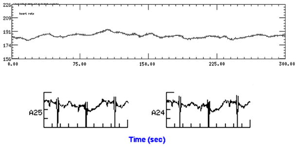
The final common pathway to death in all of us is an arrhythmia, yet we still know far too little about the contribution of conduction abnormalities and arrhythmias to the compromised states of the human fetus. At no other time in the human life cycle is the human being at more risk of unexplained and unexpected death than during the prenatal period. The risk of sudden death from 20-40 weeks gestation is 6-12 deaths/1000 fetuses/year. This is equal to, and in some ethnic groups HIGHER than, the risk of death in the adult population with known coronary artery disease over the same time frame (6-12 deaths/1000 patients/year). Because only a small percentage of the United States population is pregnant each year, because fetal demise is not often acknowledged through public displays such as funerals, and finally because fetal death is culturally accepted to a much greater extent than it should be, this critically important area of women's healthcare has not had the technological advances that have been seen in adult cardiac intensive care and other areas of medicine. Fetal cardiac deaths may be preventable and the diseases that lead to these deaths are often treatable, especially if the sophistication of our modern ICU's could somehow be translated to the prenatal monitoring arena. This review article will outline recent advances in evaluating fetal electrophysiology, helping the perinatologist to better understand the nuances of fetal arrhythmias.


Figures

Fig. 1
Fig. 1
Ectopy. The left panel shows a three-lead rhythm strip of PVCs marked by arrows. Note that no early P wave precedes these beats. The three subsequent panels show PACs conducted with a normal-appearing QRS and an aberrant QRS and not conducted through the AV node (arrows) (note the P waves).
Fig. 2
Fig. 2
Complex ventricular arrhythmias related to myocarditis, including couplets and nonsustained VT first noted by echo/Doppler (top tracing), in a 32-week gestation fetus evaluated by fMCG (middle tracing) and confirmed by postnatal monitoring (lower tracing).
Fig. 3
Fig. 3
Long QT syndrome. Two-lead fetal magnetocardiogram shows a markedly prolonged QT interval (lines). The P waves are noted by the arrows. This fetus had second-degree AVB.
Fig. 4
Fig. 4
Fetal heart rate trends demonstrate fetal rates frequently falling to approximately 75 beats/min. The lower two tracings show the fetal and composite maternal/fetal fMCG tracings during initially sinus rhythm then fetal BAB. Arrows depict the P waves.
Fig. 5
Fig. 5
This 17-week gestation fetus had evidence of clinical thyrotoxicosis with heart rates in the range of 180 to 192 beats/min. Note the beat-to-beat nonspecific T-wave changes in the lower tracing (signal averaged). This is consistent with T-wave alternans; however, it is not known whether T-wave alternans is an abnormal finding at this gestation and at this heart rate.
Fig. 6
Fig. 6
A slow re-entrant tachycardia at approximately 209 beats/min. The top tracing shows periods of atrial ectopy (saw tooth appearance) followed by SVT (solid line). The actocardiogram is shown on the bottom tracing. SVT onset is often linked to fetal movement, as shown by the minor deflections in the lower tracing.
Fig. 7
Fig. 7
Initiation (top tracing) and termination (bottom tracing) of SVT from two fetuses. The top tracing shows a slow SVT with a long ventriculoatrial interval at approximately 200 beats/min, and the bottom tracing shows a rapid SVT with a shorter ventriculoatrial interval.
Fig. 8
Fig. 8
Atrial flutter. The top tracing shows atrial flutter with 2:1 block. The atrial rate is 460 beats/min, and the ventricular rate is 230 beats/min. Flutter waves are marked with arrows. The QRS almost obscures the P wave. In a different patient in the lower two tracings, the administration of adenosine 100 μg/kg allows easy recognition of the rapid atrial flutter/fibrillation in this infant but does not terminate the tachycardia. Before adenosine, this rhythm could be mistaken for SVT. For this reason, during infusion of adenosine, continuous “real-time” paper recordings of the rhythm should be obtained.
Fig. 9
Fig. 9
Fetal LQTS with transient complete AVB, marked QT prolongation, and T-wave alternans.
Fig. 10
Fig. 10
Fetal heart rate and movement trends over 5 minutes in a fetus with CAVB and congenital heart disease. Note the flat heart rate tracing. The fetus requires postnatal pacemaker insertion. On the bottom tracing, in addition to the third-degree AVB, the corrected QT interval is also prolonged at 0.59 sec.
Fig. 11
Fig. 11
SVT termination. SVT is present initially. After administration of adenosine 100 μg/kg by rapid intravenous administration, the SVT terminates and there is a sinus pause caused by the vagal effects of the drug. The terminations always should be recorded, because sometimes the presence of Wolff-Parkinson-White pre-excitation is only seen transiently. Note here the short PR interval and delta wave on the two beats after termination.
Fig. 12
Fig. 12
Chaotic (multifocal) atrial tachycardia. Note the changing patterns of atrial rhythm on the two-lead rhythm strips. Chaotic atrial tachycardia often varies in rate and rhythm, with some periods resembling atrial flutter and other periods resembling complex atrial ectopy. Some sinus rhythm also can be noted (top tracing, right side).
Fig. 13
Fig. 13
Accelerated ventricular rhythm. Three-lead rhythm strip recorded at differing paper speeds shows the infant transitioning from sinus rhythm to accelerated ventricular rhythm on multiple occasions. Note the changing QRS morphology with wide QRS. The heart rate does not vary much with the change in rhythm.
Fig. 14
Fig. 14
Ventricular tachycardia. In the infant, ventricular tachycardia can be mistaken for supraventricular tachycardia, because the QRS is often only slightly wider than normal. The normal QRS duration in an infant is less than 0.08 seconds. Note in lead V1 that there is AV dissociation during tachycardia (arrows depict the P waves). This infant had an LV tumor.
Fig. 15
Fig. 15
Third-degree AVB. Twelve-lead ECG showing third-degree CAVB. The atrial rate is 166 beats/min, whereas the ventricular rate is only 48 beats/min. Note the AV dissociation with P waves (arrows) marching through the onset of the QRS. Also note that the QT is prolonged.

References

    1. Ferrer PL. Fetal arrhythmias. In: Deal B, Wolff GS, Gelband H, editors. Current concepts in diagnosis and treatment of arrhythmias in infants and children. Armonk (NY): Futura Publishing Company, Inc; 1998. p. 17.
    1. Strasburger JF. Fetal arrhythmias. Prog Pediatr Cardiol. 2000;11:1. - PubMed
    1. Simpson J. Fetal arrhythmias. In: Allen L, Hornberger LK, Sharland G, editors. Textbook of fetal cardiology. London: Greenwich Medical Media, Limited; 2000. p. 421.
    1. Cuneo BF. Outcome of fetal cardiac defects. Curr Opin Pediatr. 2006;18:490. - PubMed
    1. Cuneo BF, Strasburger JF, Wakai RT, et al. Conduction system disease in fetuses evaluated for irregular cardiac rhythm. Fetal Diagn Ther. 2006;21:307. - PubMed

Substances