

Sequence 168 to 177 of interphotoreceptor retinoid-binding protein (IRBP) is an antigenic epitope for autoreactive CD8 T cells in the B10RIII mouse
- PMID: 18063114
- PMCID: PMC2557023
- DOI: 10.1016/j.jneuroim.2007.10.016
Sequence 168 to 177 of interphotoreceptor retinoid-binding protein (IRBP) is an antigenic epitope for autoreactive CD8 T cells in the B10RIII mouse
Abstract
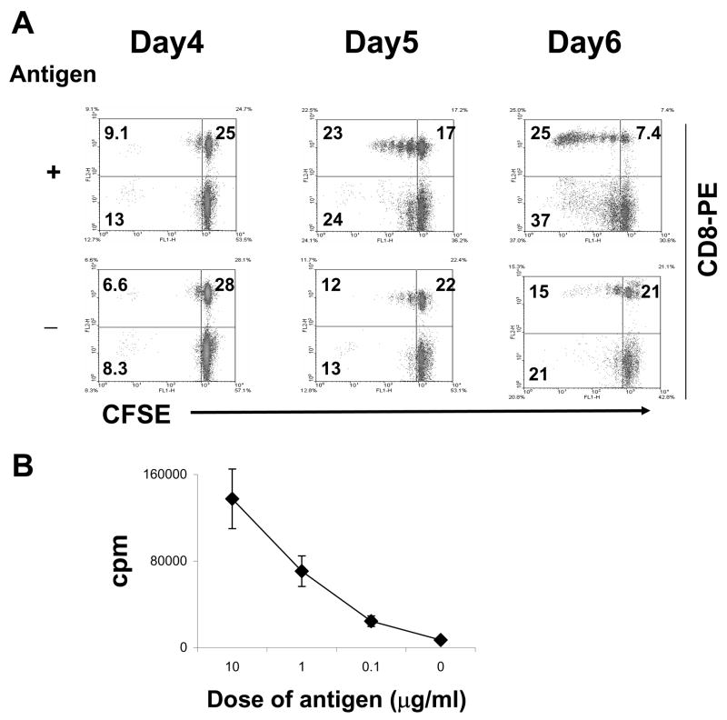
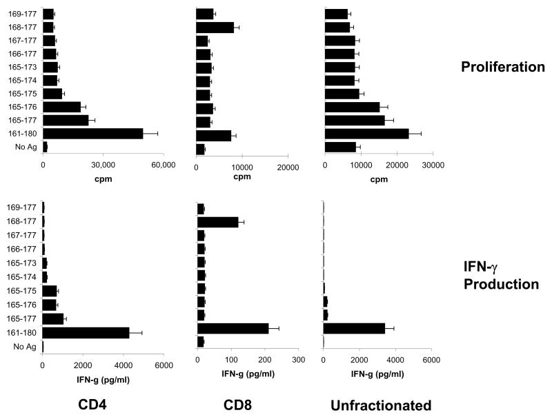
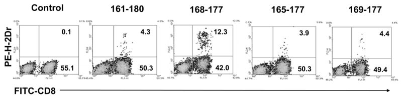
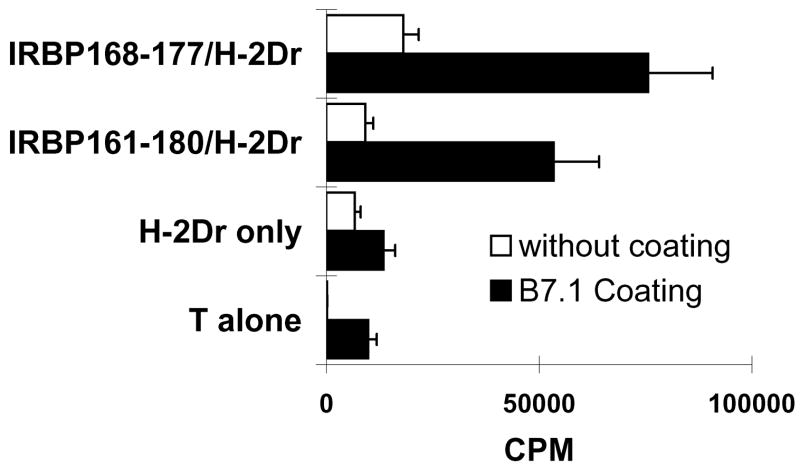
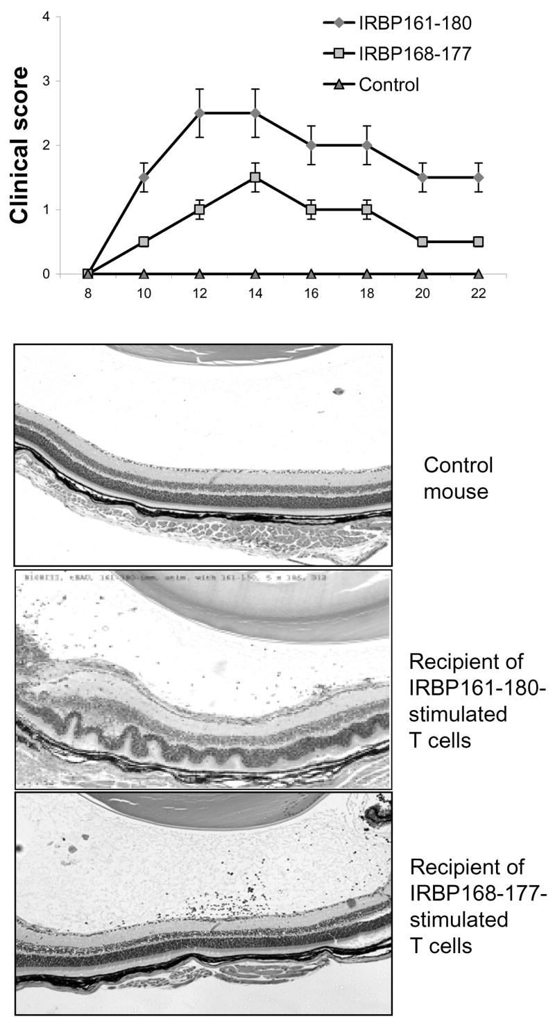
We previously demonstrated that a significant proportion of interphotoreceptor retinoid-binding protein (IRBP)-specific uveitogenic T cells in the C57BL/6 mouse and Lewis rat express CD8. The aims of this study were to determine whether some of the IRBP-specific T cells in the B10RIII mouse also express CD8 and whether CD8 and CD4 IRBP-specific T cells in the B10RIII mouse recognize a different or the same antigenic epitope. Our results show that autoreactive CD8 T cells were abundant in B10RIII mice immunized with the uveitogenic peptide IRBP161-180. Using multimers of recombinant H-2D(r) molecules, we also showed that the binding of the H-2D(r) fusion protein to IRBP161-180-expanded CD8 T cells was dependent on the peptide complexed with the recombinant molecules. The use of a panel of truncated peptides showed that the truncated 10-mer peptide, IRBP168-177, retained the ability to bind to, and stimulate, IRBP161-180-specific CD8 T cells after complexing with a dimeric MHC class I (H-2D(r)) molecule. Finally, adoptive transfer of IRBP161-180-specific T cells stimulated with IRBP168-177 consistently induced mild, but significant, EAU in naïve B10RIII mice.
Figures
Similar articles
-
A shared epitope of the interphotoreceptor retinoid-binding protein recognized by the CD4+ and CD8+ autoreactive T cells.J Immunol. 2005 Aug 1;175(3):1851-7. doi: 10.4049/jimmunol.175.3.1851. J Immunol. 2005. PMID: 16034128 Free PMC article.
-
Repertoire analysis and new pathogenic epitopes of IRBP in C57BL/6 (H-2b) and B10.RIII (H-2r) mice.Invest Ophthalmol Vis Sci. 2008 May;49(5):1946-56. doi: 10.1167/iovs.07-0868. Invest Ophthalmol Vis Sci. 2008. PMID: 18436827 Free PMC article.
-
In vitro activation of CD8 interphotoreceptor retinoid-binding protein-specific T cells requires not only antigenic stimulation but also exogenous growth factors.J Immunol. 2006 Apr 15;176(8):5006-14. doi: 10.4049/jimmunol.176.8.5006. J Immunol. 2006. PMID: 16585597
-
Minimally activated CD8 autoreactive T cells specific for IRBP express a high level of Foxp3 and are functionally suppressive.Invest Ophthalmol Vis Sci. 2007 May;48(5):2178-84. doi: 10.1167/iovs.06-1189. Invest Ophthalmol Vis Sci. 2007. PMID: 17460277 Free PMC article.
-
Identification of a new epitope of human IRBP that induces autoimmune uveoretinitis in mice of the H-2b haplotype.Invest Ophthalmol Vis Sci. 2000 Jan;41(1):127-31. Invest Ophthalmol Vis Sci. 2000. PMID: 10634611
Cited by
-
Biologic agents in experimental autoimmune uveitis.Int Ophthalmol. 2014 Feb;34(1):145-56. doi: 10.1007/s10792-013-9756-0. Epub 2013 Mar 14. Int Ophthalmol. 2014. PMID: 23494482 Review.
-
Anti-inflammatory role of IL-17 in experimental autoimmune uveitis.J Immunol. 2009 Mar 1;182(5):3183-90. doi: 10.4049/jimmunol.0802487. J Immunol. 2009. PMID: 19234216 Free PMC article.
-
Regulation of CD8(+) T Cell Responses to Retinal Antigen by Local FoxP3(+) Regulatory T Cells.Front Immunol. 2012 Jun 21;3:166. doi: 10.3389/fimmu.2012.00166. eCollection 2012. Front Immunol. 2012. PMID: 22737153 Free PMC article.
-
The role of the adaptive immune system and T cell dysfunction in neurodegenerative diseases.J Neuroinflammation. 2022 Oct 8;19(1):251. doi: 10.1186/s12974-022-02605-9. J Neuroinflammation. 2022. PMID: 36209107 Free PMC article. Review.
-
New perspectives on effector mechanisms in uveitis.Semin Immunopathol. 2008 Apr;30(2):135-43. doi: 10.1007/s00281-008-0108-5. Epub 2008 Mar 4. Semin Immunopathol. 2008. PMID: 18317764 Free PMC article. Review.
References
-
- Avichezer D, Chan CC, Silver PB, Wiggert B, Caspi RR. Residues 1-20 of IRBP and whole IRBP elicit different uveitogenic and immunological responses in interferon gamma deficient mice. Experimental Eye Research. 2000a;71:111–118. - PubMed
-
- Avichezer D, Silver PB, Chan CC, Wiggert B, Caspi RR. Identification of a new epitope of human IRBP that induces autoimmune uveoretinitis in mice of the H-2b haplotype. Invest Ophthalmol Vis Sci. 2000b;41:127–131. - PubMed
-
- Biddison WE, Cruikshank WW, Center DM, Pelfrey CM, Taub DD, Turner RV. CD8+ myelin peptide-specific T cells can chemoattract CD4+ myelin peptide-specific T cells: importance of IFN-inducible protein 10. J Immunol. 1998;160:444–448. - PubMed
-
- Fox GM, Kuwabara T, Wiggert B, Redmond TM, Hess HH, Chader GJ, Gery I. Experimental autoimmune uveoretinitis (EAU) induced by retinal interphotoreceptor retinoid-binding protein (IRBP): differences between EAU induced by IRBP and by S-antigen. Clin Immunol Immunopathol. 1987;43:256–264. - PubMed
-
- Gregerson DS, Obritsch WF, Fling SP, Cameron JD. S-antigen-specific rat T cell lines recognize peptide fragments of S-antigen and mediate experimental autoimmune uveoretinitis and pinealitis. J Immunol. 1986;136:2875–2882. - PubMed
Publication types
MeSH terms
Substances
Grants and funding
LinkOut - more resources
Full Text Sources
Research Materials
Miscellaneous
