. 2007 Dec 17;25(52):8687-701.
doi: 10.1016/j.vaccine.2007.10.030. Epub 2007 Nov 5.

Oral immunization of rhesus macaques with adenoviral HIV vaccines using enteric-coated capsules

Affiliations

Oral immunization of rhesus macaques with adenoviral HIV vaccines using enteric-coated capsules

George T Mercier et al. Vaccine. .

Abstract

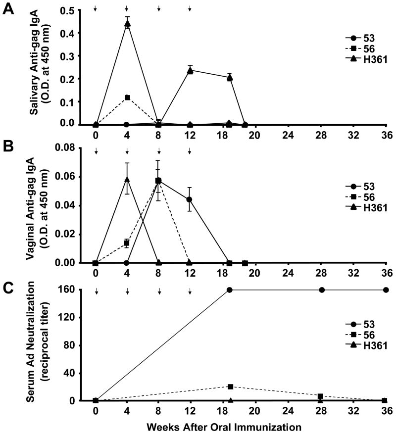
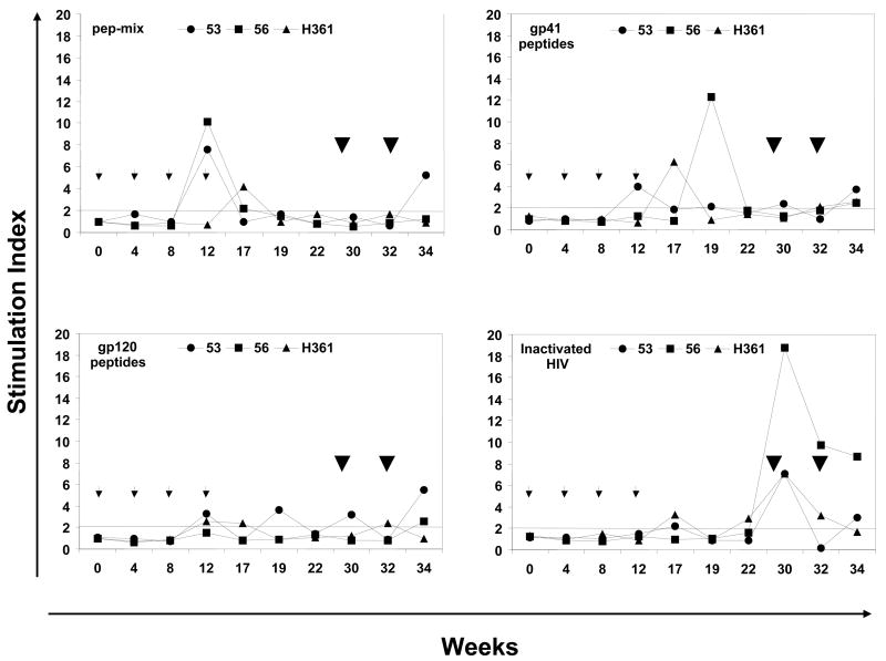
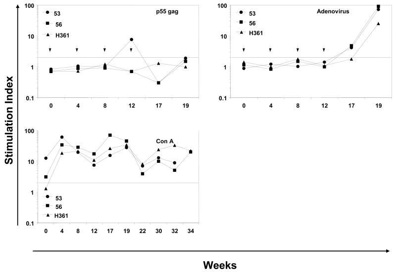
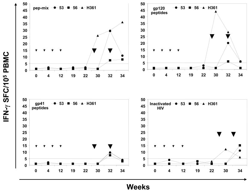
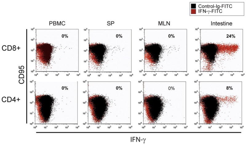
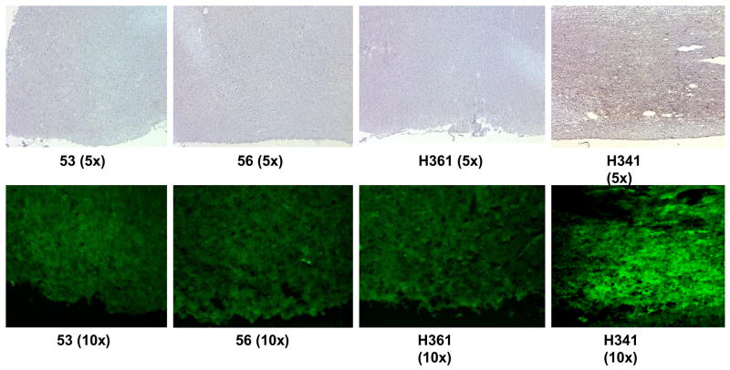
Targeted delivery of vaccine candidates to the gastrointestinal (GI) tract holds potential for mucosal immunization, particularly against mucosal pathogens like the human immunodeficiency virus (HIV). Among the different strategies for achieving targeted release in the GI tract, namely the small intestine, pH sensitive enteric coating polymers have been shown to protect solid oral dosage forms from the harsh digestive environment of the stomach and dissolve relatively rapidly in the small intestine by taking advantage of the luminal pH gradient. We developed an enteric polymethacrylate formulation for coating hydroxy-propyl-methyl-cellulose (HPMC) capsules containing lyophilized Adenoviral type 5 (Ad5) vectors expressing HIV-1 gag and a string of six highly-conserved HIV-1 envelope peptides representing broadly cross-reactive CD4(+) and CD8(+) T cell epitopes. Oral immunization of rhesus macaques with these capsules primed antigen-specific mucosal and systemic immune responses and subsequent intranasal delivery of the envelope peptide cocktail using a mutant cholera toxin adjuvant boosted cellular immune responses including, antigen-specific intracellular IFN-gamma-producing CD4(+) and CD8(+) effector memory T cells in the intestine. These results suggest that the combination of oral adenoviral vector priming followed by intranasal protein/peptide boosting may be an effective mucosal HIV vaccination strategy for targeting viral antigens to the GI tract and priming systemic and mucosal immunity.


Figures

Figure 1
Figure 1
Stability of Lyophilized Ad Vectors. Enteric-coated capsules containing lyophilized Ad vectors were immediately stored in a dessicator at 4ºC and assayed for luciferase activity on HeLa cells upon reconstitution after storage of 24 hr., 48hr., and weekly for four weeks. Percent activity was determined by comparing to the luciferase activity of infection by the same number of particles of a freshly-diluted stability control vector as was used for lyophilization. Results are presented as the means of samples prepared in triplicate, and error bars indicate standard deviations.
Figure 2
Figure 2
Enteric-coated HPMC capsule dissolution curve. Coated HPMC capsules with 13.2% w/w total solid polymer dispersion formulation of 4:1 Eudragit® L100 and Eudragit® S100 were subjected to dissolution test at 37ºC in 0.05 M sodium phosphate buffers of varying pH for the indicated times and the supernatant was assayed for A) dye release by spectrophotometry or B) virus release by real-time PCR. Results are presented as the means of samples performed in triplicate, and error bars represent the standard deviation.
Figure 3
Figure 3
Antigen-specific humoral immune responses. A and B, Antibody responses against HIV-1 gag in mucosal samples. C, Serum neutralizing responses against adenovirus. ELISA plates were coated with HIV-1 gag and 1:2 dilutions of saliva (A) and vaginal washes (B) were reacted. After reaction with secondary anti-IgA HRP conjugate and subsequent colorimetric substrate, data were presented as the O.D. values measured at 450 nm. Error bars indicate standard deviations of three measurements taken per time point. Statistical significance was verified for each time point against the background using a Student’s paired t test and a p value < 0.05. C) Serum samples take at the indicated times were incubated with Ad5 expressing luciferase for 1 hour at 37°C prior to addition to 293 cells at 250 viral particles per cel. 24 hours later, luciferase activity was measured and gene delivery was compared to untreated Ad5 vector. Data is expressed as geometric mean titers that reduced Ad luciferase activity 50%.
Figure 4
Figure 4
Antigen-specific cellular immune responses. The proliferative responses in terms of [3H] thymidine incorporation and IFN-γ producing cells by the ELISPOT method were determined using PBMC samples from rhesus macaques immunized by oral priming with enteric-coated capsules delivering Ad-gag and Ad-env-peptide and intranasal boosting with synthetic HIV-1 envelope peptides and CT2*, the mutant cholera toxin adjuvant. (A) Systemic T cell responses using PBMC were determined in terms of proliferation in response to mixtures of all six HIV-1 envelope peptides (pep-mix), two gp41 peptides, four gp120 peptides, and heat-inactivated HIV-1IIIB preparation. The fold-increases in proliferation responses to the different antigens were calculated by comparing the values from cells in the culture medium. (B) Proliferation responses of PBMC were determined in response to recombinant HIV-1 gag protein (p55 gag), heat-inactivated Ad5, and Con A. (C) ELISPOT analyses for antigen-specific IFN-γ-producing cells for 105 input PBMC in each monkey, in terms of spot-forming-cells (SFC/105 PBMC) at the indicated times after oral Ad vector mediated priming and intranasal boosting with peptide-cocktail (6 peptides, 100 μg each/dose) mixed with mutant cholera toxin, CT2* (10 μg/dose). The horizontal line in each panel indicates the cut-off value for positive response (SI of 2.0 for the proliferation assay and SFC of 5 for the ELISPOT assay).
Figure 4
Figure 4
Antigen-specific cellular immune responses. The proliferative responses in terms of [3H] thymidine incorporation and IFN-γ producing cells by the ELISPOT method were determined using PBMC samples from rhesus macaques immunized by oral priming with enteric-coated capsules delivering Ad-gag and Ad-env-peptide and intranasal boosting with synthetic HIV-1 envelope peptides and CT2*, the mutant cholera toxin adjuvant. (A) Systemic T cell responses using PBMC were determined in terms of proliferation in response to mixtures of all six HIV-1 envelope peptides (pep-mix), two gp41 peptides, four gp120 peptides, and heat-inactivated HIV-1IIIB preparation. The fold-increases in proliferation responses to the different antigens were calculated by comparing the values from cells in the culture medium. (B) Proliferation responses of PBMC were determined in response to recombinant HIV-1 gag protein (p55 gag), heat-inactivated Ad5, and Con A. (C) ELISPOT analyses for antigen-specific IFN-γ-producing cells for 105 input PBMC in each monkey, in terms of spot-forming-cells (SFC/105 PBMC) at the indicated times after oral Ad vector mediated priming and intranasal boosting with peptide-cocktail (6 peptides, 100 μg each/dose) mixed with mutant cholera toxin, CT2* (10 μg/dose). The horizontal line in each panel indicates the cut-off value for positive response (SI of 2.0 for the proliferation assay and SFC of 5 for the ELISPOT assay).
Figure 5
Figure 5
Antigen-specific intracellular IFN-γ production. Cells isolated from various tissues at necropsy of macaque 53 were used for determining intracellular IFN-γ producing T cells by immunofluorescence staining after stimulation with the HIV-1 envelope peptide mixture (pep-mix), cell fixation and permeabilization as described in the methods section. To define T cell subsets, lymphocytes were stained for cell surface CD4, CD8, and CD95, the later to distinguish memory-phenotype. Among the CD95+ T cells, IFN-γ + CD8+ and CD4+ T cells were detected in the intestinal lymphocytes, but not in peripheral blood, spleen, and lymph nodes.
Figure 6
Figure 6
Detection of NGF-b1 expression in the olfactory bulbs of rhesus macaques immunized by the intranasal route with the HIV-1 envelope peptide cocktail (pep-mix) using either the mucosal adjuvant mutant cholera toxin CT2* (the first three panels on the left-side indicated by monkey numbers, 53, 56, and H361) or native cholera toxin (nCT, right-side panel indicated by monkey number H341). Frozen tissue sections were subjected to anti-NGF-b1 antibody immunohistochemistry staining (upper panels) and immunofluoresence straining (lower panels) by reacting with avidin-biotin conjugate, followed by incubation with HRP-conjugated streptavidin or streptavidin-Alexa Fluor 488.

References

    1. Shiver JW, Emini EA. Recent advances in the development of HIV-1 vaccines using replication-incompetent adenovirus vectors. Annu Rev Med. 2004;55:355–372. - PubMed
    1. Belyakov IM, Derby MA, Ahlers JD, et al. Mucosal immunization with HIV-1 peptide vaccine induces mucosal and systemic cytotoxic T lymphocytes and protective immunity in mice against intrarectal recombinant HIV-vaccinia challenge. Proc Natl Acad Sci U S A. 1998;95(4):1709–1714. - PMC - PubMed
    1. Belyakov IM, Ahlers JD, Brandwein BY, et al. The importance of local mucosal HIV-specific CD8(+) cytotoxic T lymphocytes for resistance to mucosal viral transmission in mice and enhancement of resistance by local administration of IL-12. J Clin Invest. 1998;102(12):2072–2081. - PMC - PubMed
    1. Gallichan WS, Rosenthal KL. Long-term immunity and protection against herpes simplex virus type 2 in the murine female genital tract after mucosal but not systemic immunization. J Infect Dis. 1998;177(5):1155–1161. - PubMed
    1. Gallichan WS, Rosenthal KL. Long-lived cytotoxic T lymphocyte memory in mucosal tissues after mucosal but not systemic immunization. J Exp Med. 1996;184(5):1879–1890. - PMC - PubMed

Publication types

MeSH terms