

Advances in the diagnosis and therapy of paroxysmal nocturnal hemoglobinuria
- PMID: 18063459
- PMCID: PMC2290854
- DOI: 10.1016/j.blre.2007.10.002
Advances in the diagnosis and therapy of paroxysmal nocturnal hemoglobinuria
Abstract
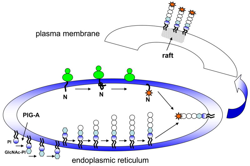
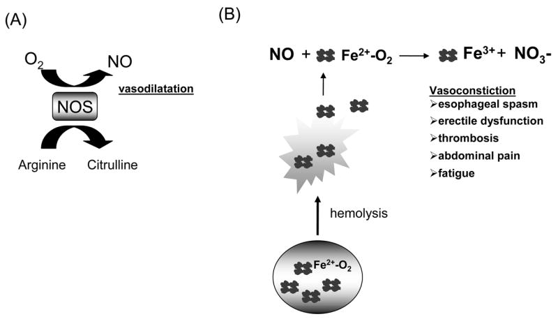
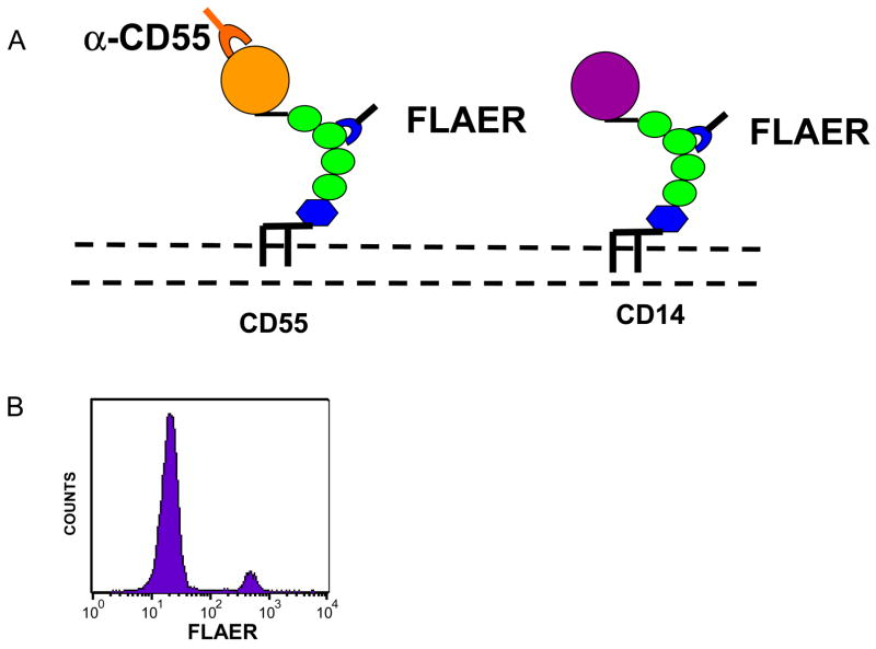
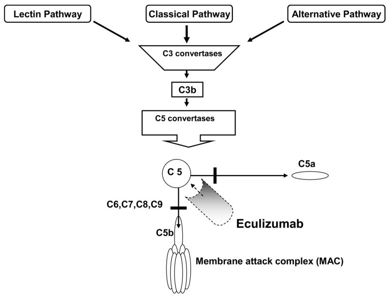
PNH is an uncommon acquired hemolytic anemia that often manifests with hemoglobinuria, abdominal pain, smooth muscle dystonias, fatigue, and thrombosis. The disease results from the expansion of hematopoietic stem cells harboring a mutation in a gene, PIG-A, that is required for the biosynthesis of a lipid moiety, glycosylphosphatidylinositol (GPI), that attaches dozens of different proteins to the cell surface. Thus, PNH cells are deficient in cell surface GPI anchored proteins; this deficiency on erythrocytes leads to intravascular hemolysis since certain GPI anchored proteins normally function as complement regulators. Free hemoglobin released from intravascular hemolysis leads to circulating nitric oxide depletion and is responsible for many of the clinical manifestations of PNH, including fatigue, erectile dysfunction, esophageal spasm, and thrombosis. Interestingly, rare PIG-A mutations can be found in virtually all healthy control subjects leading to speculation that PIG-A mutations in hematopoietic stem cells are common benign events. However, recent data reveals that most of these mutations in healthy controls are not derived from stem cells. The recently FDA approved complement inhibitor eculizumab has been shown to decrease hemolysis, decrease erythrocyte transfusion requirements, decrease the risk for thrombosis and improve quality of life for PNH patients.
Figures
Similar articles
-
New insights into paroxysmal nocturnal hemoglobinuria.Hematology. 2007 Oct;12(5):371-6. doi: 10.1080/10245330701562634. Hematology. 2007. PMID: 17852463 Review.
-
New insights into paroxysmal nocturnal hemoglobinuria.Hematology Am Soc Hematol Educ Program. 2006:24-8, 516. doi: 10.1182/asheducation-2006.1.24. Hematology Am Soc Hematol Educ Program. 2006. PMID: 17124035 Review.
-
[Eculizumab in paroxysmal nocturnal hemoglobinuria].Med Sci (Paris). 2009 Dec;25(12):1126-9. doi: 10.1051/medsci/200925121126. Med Sci (Paris). 2009. PMID: 20035691 Review. French.
-
Paroxysmal nocturnal hemoglobinuria from bench to bedside.Clin Transl Sci. 2011 Jun;4(3):219-24. doi: 10.1111/j.1752-8062.2011.00262.x. Clin Transl Sci. 2011. PMID: 21707954 Free PMC article. Review.
-
Improvement in the symptoms of smooth muscle dystonia during eculizumab therapy in paroxysmal nocturnal hemoglobinuria.Haematologica. 2005 Dec;90(12 Suppl):ECR40. Haematologica. 2005. PMID: 16464755
Cited by
-
[Chinese expert consensus on paroxysmal nocturnal hemoglobinuria detection via flow cytometry (2021)].Zhonghua Xue Ye Xue Za Zhi. 2021 Apr 14;42(4):281-287. doi: 10.3760/cma.j.issn.0253-2727.2021.04.003. Zhonghua Xue Ye Xue Za Zhi. 2021. PMID: 33979971 Free PMC article. Chinese. No abstract available.
-
Beyond the Role of CD55 as a Complement Component.Immune Netw. 2018 Feb 20;18(1):e11. doi: 10.4110/in.2018.18.e11. eCollection 2018 Feb. Immune Netw. 2018. PMID: 29503741 Free PMC article. Review.
-
Predictive Factors of Mortality in Population of Patients with Paroxysmal Nocturnal Hemoglobinuria (PNH): Results from a Korean PNH Registry.J Korean Med Sci. 2016 Feb;31(2):214-21. doi: 10.3346/jkms.2016.31.2.214. Epub 2016 Jan 26. J Korean Med Sci. 2016. PMID: 26839475 Free PMC article.
-
Paroxysmal nocturnal hemoglobinuria: role of the complement system, pathogenesis, and pathophysiology.J Manag Care Spec Pharm. 2020 Dec;26(12-b Suppl):S3-S8. doi: 10.18553/jmcp.2020.26.12-b.s3. J Manag Care Spec Pharm. 2020. PMID: 33356782 Free PMC article. Review.
-
Targeted inhibition of complement using complement receptor 2-conjugated inhibitors attenuates EAE.Neurosci Lett. 2012 Nov 30;531(1):35-9. doi: 10.1016/j.neulet.2012.10.012. Epub 2012 Oct 16. Neurosci Lett. 2012. PMID: 23079547 Free PMC article.
References
-
- Brodsky RA. Paroxysmal nocturnal hemoglobinuria. In: Hoffman R, Benz EJ Jr, Shattil SJ, et al., editors. Hematology: Basic Principles and Practice. Philadelphia: E; 2005. pp. 419–427.
-
- Strubing P. Paroxysmale Haemoglobinurie. Deutsche Medicinische Wochenschrift. 1882;8:1–16.
-
- Enneking J. Eine neue form intermittierender haemoglobinurie (Haemoglobinuria paroxysmalis nocturia) Klinische Wochenschrift. 1928;7:2045.
-
- Ham T. Chronic hemolytic anemia with paroxysmal nocturnal hemoglobinuria. A study of the mechanism of hemolysisin relation to acid-base equilibrium. New England Journal of Medicine. 1937;217:915–917.
-
- Pillemer L, Blum L, Lepow I, et al. The properdin system and immunity: Demonstration and isolation of a new serum protein, properdin, and its role in immune phenomena. Science. 1954;120:279–285. - PubMed
Publication types
MeSH terms
Substances
Grants and funding
LinkOut - more resources
Full Text Sources
Other Literature Sources
