. 2008 Feb;36(2):616-28.
doi: 10.1093/nar/gkm556. Epub 2007 Dec 5.

Nuclear RNA export factor 7 is localized in processing bodies and neuronal RNA granules through interactions with shuttling hnRNPs

Affiliations

Nuclear RNA export factor 7 is localized in processing bodies and neuronal RNA granules through interactions with shuttling hnRNPs

Jun Katahira et al. Nucleic Acids Res. 2008 Feb.

Abstract

The nuclear RNA export factor (NXF) family proteins have been implicated in various aspects of post-transcriptional gene expression. This study shows that mouse NXF7 exhibits heterologous localization, i.e. NXF7 associates with translating ribosomes, stress granules (SGs) and processing bodies (P-bodies), the latter two of which are believed to be cytoplasmic sites of storage, degradation and/or sorting of mRNAs. By yeast two-hybrid screening, a series of heterogeneous nuclear ribonucleoproteins (hnRNPs) were identified as possible binding partners for NXF7. Among them, hnRNP A3, which is believed to be involved in translational control and/or cytoplasmic localization of certain mRNAs, formed a stable complex with NXF7 in vitro. Although hnRNP A3 was not associated with translating ribosomes, it was co-localized with NXF7 in P-bodies. After exposing to oxidative stress, NXF7 trans-localized to SGs, whereas hnRNP A3 did not. In differentiated neuroblastoma Neuro2a cells, NXF7 was co-localized with hnRNP A3 in cell body and neurites. The amino terminal half of NXF7, which was required for stable complex formation with hnRNP A3, coincided with the region required for localization in both P-bodies and neuronal RNA granules. These findings suggest that NXF7 plays a role in sorting, transport and/or storage of mRNAs through interactions with hnRNP A3.


Figures
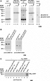
Figure 1.
Figure 1.
NXF7 binds a series of shuttling hnRNPs. (A) GST (lanes 1 to 3), GST-Tap (aa 96–371; lanes 4 to 6), GST-NXF7 (full length; lanes 7 to 9), and GST-NXF7 (aa 99–374; lanes 10 to 12), each pre-adsorbed to glutathione Sepharose beads, were incubated for 2 h at 4°C with either buffer (lanes 1, 4, 7, 11), hnRNP A3 (lanes 2, 5, 8, 11) or hnRNP A2/B1 (lanes 3, 6, 9, 12). After washing extensively, bound proteins were eluted by boiling in SDS sample buffer and were then loaded on SDS-12% polyacrylamide gels. In lanes 13 and 14, ∼5% of input was loaded. Proteins were visualized by staining the gel with Coomassie brilliant blue. Migration positions of molecular weight markers are indicated on the left in kilo Daltons and those of hnRNP A3 and hnRNP A2/B1 are on the right side of the panel, indicated by arrowheads. Asterisks indicate the positions of GST and GST fusion proteins. (B) Same as in A, but proteins were detected by Western blot using the indicated antibodies. (C) Same as in A, but 6xHis-tagged KSRP was subjected to binding reactions. Proteins were detected by Western blot using anti-pentaHis antibody.
Figure 2.
Figure 2.
NXF7 is localized in polyribosomes containing fractions. (A) Upper panel: Cytoplasmic lysate of a mouse L929 cell line stably expressing FLAG-tagged NXF7 was prepared using Mg++-containing buffer and was fractionated over a linear 20–50% sucrose gradient, which also contained Mg++. The ribosome profile (OD254 nm trace) is depicted. Arrows indicate the positions of 40, 60 and 80 S ribosomal subunits in the gradient, while a bracket illustrates positions of polyribosomes. Total RNA and protein were isolated from each fraction. Distribution of tRNAs and rRNAs was examined by denaturing agarose gel electrophoresis followed by ethidium bromide staining of the gel. FLAG-tagged NXF7 fusion protein was detected by Western blot with an anti-FLAG antibody. Lower panel: Cytoplasmic lysate of a mouse L929 cell line stably expressing FLAG-tagged NXF7 was prepared using an EDTA-containing buffer. Fractionation was performed as above, but the gradient contained 50 mM EDTA. The positions of small and large ribosome subunits are shown by arrows in the OD254 nm trace. Distribution of RNAs and FLAG-tagged NXF7 was examined as above. (B) Upper panel: Cytoplasmic lysate of a mouse L929 cell line stably expressing FLAG-tagged NXF7 was prepared and fractionated under Mg++-containing condition as in A. Lower left panel: Cytoplasmic lysate was prepared after the cells were treated with 1 mM puromycin for 5 h. Lower right panel: Cytoplasmic lysate was prepared in the presence of RNase A (100 μg/ml). In both experiments, fractionation was performed under Mg++-containing condition. The ribosome profiles (OD254 nm trace) are depicted. The positions of 40, 60 and 80 S ribosomal subunits and polyribosomes in the gradients are indicated in each panel. FLAG-tagged NXF7 and hnRNP A3 in the gradients were detected by Western blot using anti-FLAG and anti-hnRNP A3 antibodies, respectively. A non-specific band that migrates slower than NXF7-FLAG is indicated by asterisks.
Figure 3.
Figure 3.
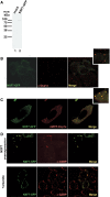
NXF7 is localized in P-bodies and SGs. (A) HeLa cells were transiently transfected with a vector harboring NXF7-GFP fusion protein (lane 2). Untransfected HeLa cells were used as negative control (lane 1). At 48 h after transfection, total cell extracts were prepared and subjected to Western blot using anti-GFP antibody. Positions of molecular weight markers are indicated on the left in kDa. (B) HeLa cells were transiently transfected with a vector harboring NXF7-GFP fusion protein. The cells were fixed 20 h after transfection and localization of Dcp1a was detected by immunofluorescence using an anti-Dcp1a antibody followed by Alexa568-labeled anti-rabbit IgG. The upper right panel is a magnified view of the area indicated by the white box in the merged image. (C) HeLa cells were transfected with vectors harboring NXF7-GFP and mRFP-Dcp1a fusion proteins. At 20 h after transfection, the cells were fixed and observed by epifluorescent microscopy. The upper right panel is a magnified view of the area indicated by the white box in the merged image. (D) Upper panels: HeLa cells expressing NXF7-GFP were fixed and subjected to immunofluorescence using an anti-G3BP antibody followed by Alexa568-labeled anti-mouse IgG. Note that in highly expressing cells, aggregated NXF7-GFP contained G3BP. Lower panels: HeLa cells expressing NXF7-GFP were exposed to oxidative stress for 1 h, fixed, and subjected to immunofluorescence as above. Note that even in the cells expressing low amounts of the fusion protein, NXF7 relocalized to SGs.
Figure 4.
Figure 4.
Identification of domains required for P-body and SG localization. (A) A schematic representation of the mutants used. The names of each domain are based on our previous reports (10). The numbers above each rectangle indicate amino acid positions. + and − signs indicate the presence or absence of mutants in P-bodies and SGs. (B) Total cell extracts prepared from HeLa cells transiently expressing each mutant were separated by SDS–PAGE and subjected to Western blot using a rabbit anti-GFP antibody. The positions of molecular weight markers are indicated on the left side in kDa. Asterisks indicate the positions of degradation products. (C) HeLa cells co-expressing mRFP-Dcp1a and the CFP-tagged mutants were observed by epifluorescent microscopy. (D) Co-localization of CFP-NXF7(1–374) and mRFP-Dcp1a in transfected HeLa cells was observed by confocal microscopy. Insets are magnified view of the areas indicated by the white boxes. (E) HeLa cells expressing CFP-tagged mutants were cultured for 1 h in the presence of 0.5 mM arsenite before fixation. Localization of G3BP was detected by immunofluorescence using mouse anti-G3BP antibody followed by Alexa568-labeled anti-mouse IgG. The cells were observed by confocal microscopy.
Figure 4.
Figure 4.
Identification of domains required for P-body and SG localization. (A) A schematic representation of the mutants used. The names of each domain are based on our previous reports (10). The numbers above each rectangle indicate amino acid positions. + and − signs indicate the presence or absence of mutants in P-bodies and SGs. (B) Total cell extracts prepared from HeLa cells transiently expressing each mutant were separated by SDS–PAGE and subjected to Western blot using a rabbit anti-GFP antibody. The positions of molecular weight markers are indicated on the left side in kDa. Asterisks indicate the positions of degradation products. (C) HeLa cells co-expressing mRFP-Dcp1a and the CFP-tagged mutants were observed by epifluorescent microscopy. (D) Co-localization of CFP-NXF7(1–374) and mRFP-Dcp1a in transfected HeLa cells was observed by confocal microscopy. Insets are magnified view of the areas indicated by the white boxes. (E) HeLa cells expressing CFP-tagged mutants were cultured for 1 h in the presence of 0.5 mM arsenite before fixation. Localization of G3BP was detected by immunofluorescence using mouse anti-G3BP antibody followed by Alexa568-labeled anti-mouse IgG. The cells were observed by confocal microscopy.
Figure 5.
Figure 5.
NXF7 is co-localized with hnRNP A3 in P-bodies. (A) HeLa cells expressing NXF7-GFP (left panels) or GFP-Dcp1a were fixed and subjected to immunofluorescence using rabbit anti-hnRNP A3 antibody. The lower left and right panels are magnified images of the areas indicated by white boxes. Localization was observed by confocal microscopy. (B) HeLa cells expressing NXF7-GFP and mRFP-Dcp1a were fixed and subjected to immunofluorescence as in A. Visualization of localization of hnRNP A3 was done using Alexa647-labeled anti-rabbit IgG. The lowest panel shows a magnified view of the area indicated by a white box in the merged image. (C) CEB-derived cells cultured on glass bottom dishes were fixed and immunostained with anti-Dcp1a and anti-NXF7 antibodies followed by Alexa568-labeled anti-rabbit and Alexa488-labeled anti-rat secondary antibodies. The cells were observed by confocal microscopy. The lower right panel shows a magnified view of the area indicated by the white box in the merged image. (D) Same as in C, but the cells were immunostained with anti-hnRNP A3 and anti-NXF7 antibodies. The cells were observed by confocal microscopy. The lower right panel shows a magnified view of the area indicated by the white box in the merged image. (E) HeLa cells expressing NXF7-GFP were cultured in the presence of 0.5 mM arsenite for 1 h. The cells were fixed and immunostained with anti-Dcp1a (upper panels) and anti-hnRNP A3 (lower panels) antibodies followed by Alexa568-labeled anti-rabbit IgG. Localization of each protein was detected by confocal microscopy.
Figure 5.
Figure 5.
NXF7 is co-localized with hnRNP A3 in P-bodies. (A) HeLa cells expressing NXF7-GFP (left panels) or GFP-Dcp1a were fixed and subjected to immunofluorescence using rabbit anti-hnRNP A3 antibody. The lower left and right panels are magnified images of the areas indicated by white boxes. Localization was observed by confocal microscopy. (B) HeLa cells expressing NXF7-GFP and mRFP-Dcp1a were fixed and subjected to immunofluorescence as in A. Visualization of localization of hnRNP A3 was done using Alexa647-labeled anti-rabbit IgG. The lowest panel shows a magnified view of the area indicated by a white box in the merged image. (C) CEB-derived cells cultured on glass bottom dishes were fixed and immunostained with anti-Dcp1a and anti-NXF7 antibodies followed by Alexa568-labeled anti-rabbit and Alexa488-labeled anti-rat secondary antibodies. The cells were observed by confocal microscopy. The lower right panel shows a magnified view of the area indicated by the white box in the merged image. (D) Same as in C, but the cells were immunostained with anti-hnRNP A3 and anti-NXF7 antibodies. The cells were observed by confocal microscopy. The lower right panel shows a magnified view of the area indicated by the white box in the merged image. (E) HeLa cells expressing NXF7-GFP were cultured in the presence of 0.5 mM arsenite for 1 h. The cells were fixed and immunostained with anti-Dcp1a (upper panels) and anti-hnRNP A3 (lower panels) antibodies followed by Alexa568-labeled anti-rabbit IgG. Localization of each protein was detected by confocal microscopy.
Figure 6.
Figure 6.
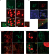
Co-localization of NXF7 and hnRNP A3 in distal sites of neurites. (A) Neuro2a cells expressing NXF7-GFP were fixed and subjected to immunofluorescence using anti-hnRNP A3 antibody. Localization was observed by confocal microscopy. Insets are magnified view of the areas indicated by the white boxes. (B) Same as in A, but immunofluorescence was performed using anti-Dcp1a antibody. Inset is a magnified view of the area indicated by the white box. (C) Neuro2a cells expressing a CFP-fusion protein containing the N+LRR domain (aa 1–374) of NXF7 was subjected to immunofluorescence using anti-hnRNP A3 antibody as in A. (D) Neuro2a cells expressing a CFP-fusion protein containing the M+C domain (aa 375–620) of NXF7 was subjected to immunofluorescence using anti-hnRNP A3 antibody as in A. (E) Neuro2a cells expressing a CFP-fusion protein containing the minimal hnRNP A3-binding domain (aa 91–374 of NXF7) was subjected to immunofluorescence using anti-hnRNP A3 antibody as in A.
Figure 7.
Figure 7.
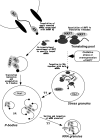
A model for the possible function of NXF7. See ‘Discussion’ section for details.

Similar articles

Cited by

References

    1. Segref A, Sharma K, Doye V, Hellwig A, Huber J, Luhrmann R, Hurt E. Mex67p, a novel factor for nuclear mRNA export, binds to both poly(A)+ RNA and nuclear pores. EMBO J. 1997;16:3256–3271. - PMC - PubMed
    1. Gruter P, Tabernero C, von Kobbe C, Schmitt C, Saavedra C, Bachi A, Wilm M, Felber BK, Izaurralde E. TAP, the human homolog of Mex67p, mediates CTE-dependent RNA export from the nucleus. Mol. Cell. 1998;1:649–659. - PubMed
    1. Katahira J, Strasser K, Podtelejnikov A, Mann M, Jung JU, Hurt E. The Mex67p-mediated nuclear mRNA export pathway is conserved from yeast to human. EMBO J. 1999;18:2593–2609. - PMC - PubMed
    1. Kang Y, Cullen BR. The human Tap protein is a nuclear mRNA export factor that contains novel RNA-binding and nucleocytoplasmic transport sequences. Genes Dev. 1999;13:1126–1139. - PMC - PubMed
    1. Herold A, Suyama M, Rodrigues JP, Braun IC, Kutay U, Carmo-Fonseca M, Bork P, Izaurralde E. TAP (NXF1) belongs to a multigene family of putative RNA export factors with a conserved modular architecture. Mol. Cell. Biol. 2000;20:8996–9008. - PMC - PubMed

Publication types

MeSH terms

Substances