. 2008 Mar;149(3):979-87.
doi: 10.1210/en.2007-1282. Epub 2007 Dec 6.

Human chorionic gonadotropin stimulates trophoblast invasion through extracellularly regulated kinase and AKT signaling

Affiliations

Human chorionic gonadotropin stimulates trophoblast invasion through extracellularly regulated kinase and AKT signaling

Johanna Prast et al. Endocrinology. 2008 Mar.

Abstract

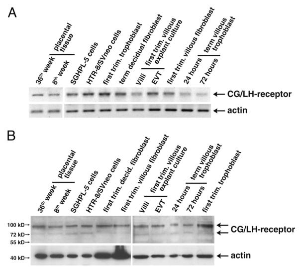
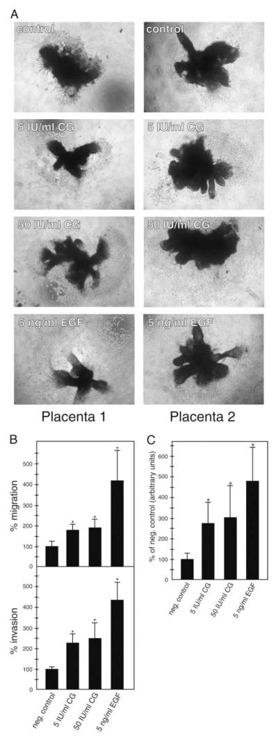
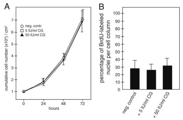
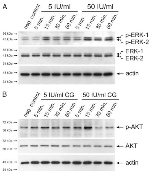
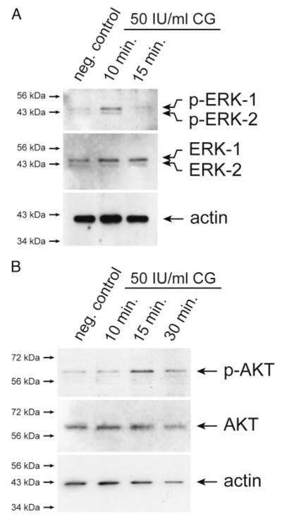
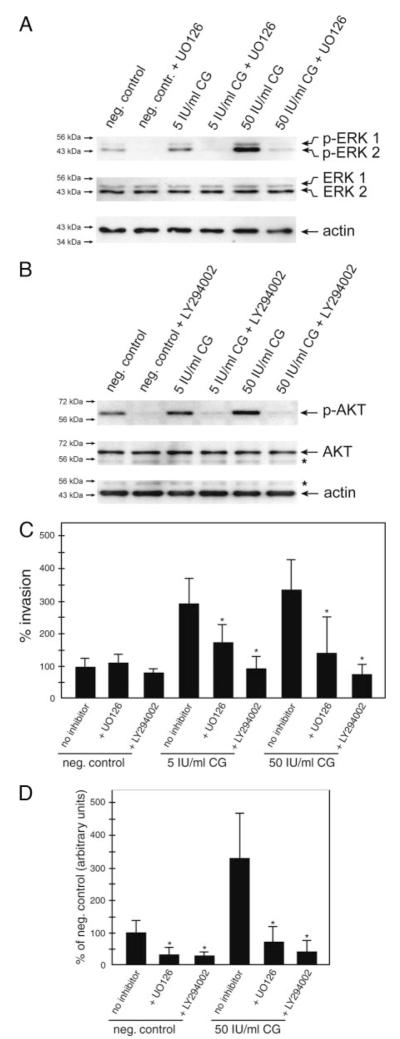
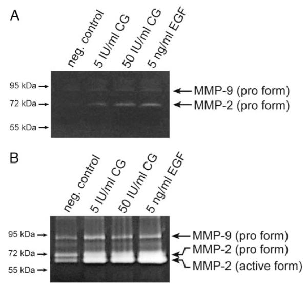
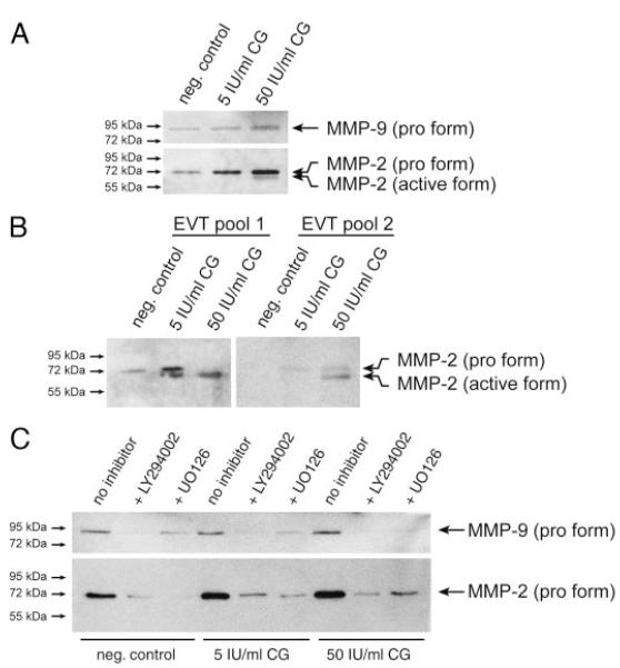
Chorionic gonadotropin (CG) is indispensable for human pregnancy because it controls implantation, decidualization, and placental development. However, its particular role in the differentiation process of invasive trophoblasts has not been fully unraveled. Here we demonstrate that the hormone promotes trophoblast invasion and migration in different trophoblast model systems. RT-PCR and Western blot analyses revealed expression of the LH/CG receptor in trophoblast cell lines and different trophoblast primary cultures. In vitro, CG increased migration and invasion of trophoblastic SGHPL-5 cells through uncoated and Matrigel-coated transwells, respectively. The hormone also increased migration of first-trimester villous explant cultures on collagen I. Proliferation of the trophoblast cell line and villous explant cultures measured by cumulative cell numbers and in situ 5-bromo-2'-deoxyuridine labeling, respectively, was unaffected by CG. Addition of the hormone activated ERK-1/2 and AKT in SGHPL-5 cells and pure, extravillous trophoblasts. Inhibition of MAPK kinase/ERK and phosphatidylinositide 3-kinase/AKT blocked phosphorylation of the kinases and attenuated CG-dependent invasion of SGHPL-5 cells. Similarly, the inhibitors decreased hormone-stimulated migration in villous explant cultures. Western blot analyses and gelatin zymography suggested that CG increased matrix metalloproteinase (MMP)-2 protein levels and activity in both culture systems. Inhibition of ERK or AKT diminished CG-induced MMP-2 expression. In summary, the data demonstrate that CG promotes trophoblast invasion and migration through activation of ERK and AKT signaling involving their downstream effector MMP-2. Because the increase of CG during the first trimester of pregnancy correlates with rising trophoblast motility, the hormone could be a critical regulator of the early invasion process.


Figures

Fig. 1
Fig. 1
Expression analyses of LH/CG receptor in placental tissues and different trophoblast model systems. RNA and protein lysates were prepared from placental tissues, first-trimester villous explant cultures, primary trophoblasts and fibroblasts, and trophoblastic cell lines. RT-PCR and Western blot analyses were done as described in Materials and Methods. Representative examples are shown. A, Semiquantitative RT-PCR analyses detecting a 508-bp LH/CG receptor fragment. β-Actin (398 bp) was used as a loading control. B, Western blot analyses using specific antibodies against LH/CG receptor. Actin was used as loading control. Specific signals are indicated by arrows; marker bands are depicted.
Fig. 2
Fig. 2
CG increases migration and invasion in different trophoblast model systems. A, Migration and invasion of SGHPL-5 cells through uncoated and Matrigel-coated transwells, respectively. Assays and stimulation with CG was performed as described in Materials and Methods. EGF was used as a positive control. Bars represent mean values of three (migration) and four (invasion) different experiments performed in duplicates; error bars indicate SD. Mean value of untreated cultures (negative control) was arbitrarily set to 100%. *, P < 0.05. B, CG-induced migration in first-trimester villous explant cultures cultivated on collagen I. Preparation of organ cultures and CG treatment was done as mentioned above. Representative examples of explants isolated from two different placentae were chosen and digitally photographed (10-fold magnification). C, Quantification of the area of outgrowth in CG-treated explant cultures. Mean values ± SD of 24 explants per condition derived from three different placentae are depicted. *, P < 0.05.
Fig. 3
Fig. 3
Proliferation of SGHPL-5 cells and villous explant cultures in the absence or presence of CG. A, Determination of cumulative cell numbers. SGHPL-5 cells (n = 3) were cultivated up to 72 h with different doses of CG and counted as mentioned in Materials and Methods. Mean values of three different experiments performed in duplicates are shown; error bars depict SD. *, Significant change (P < 0.05) at 72 h. B, Proliferation in villous explant cultures. BrdU labeling, CG stimulation, and determination of number of BrdU-labeled nuclei in cell columns were done as described above. Mean values (percentage of BrdU labeled nuclei) ± SD of three different placentae are depicted. *, P < 0.05.
Fig. 4
Fig. 4
Western blot analyses showing CG-dependent phosphorylation of ERK and AKT in SGHPL-5 cells. Serum-free cultures were treated with either 5 or 50 IU/ml CG for indicated time periods. Preparation of protein lysates and Western blot analyses were performed as described in Materials and Methods. Marker bands (kilodaltons) are depicted on the left side. Actin (42 kDa) was used as a loading control. Representative examples of n = 3 are shown. A, CG-stimulated activation of ERK. Specific signals of phosphorylated ERK-1 (44 kDa) and ERK-2 (42 kDa) and total ERK-1/2 are indicated by arrows. B, Phosphorylation of AKT after CG treatment. Arrows indicate specific signals of phosphorylated and total AKT protein (60 kDa), respectively.
Fig. 5
Fig. 5
Western blot analyses demonstrating CG-stimulated phosphorylation of ERK and AKT in pure, EVTs. Preparation of EVT, CG stimulation, and detection of ERK and AKT phosphorylation were performed as described in Materials and Methods. Marker bands (kilodaltons) are depicted on the left side. Actin (42 kDa) was used to evaluate protein loading. Representative examples are shown. A, Phosphorylation of ERK. Specific bands of p-ERK-1/2 and total ERK-1/2 are indicated. B, Activation of AKT. Arrows specify signals of phosphorylated and total AKT protein.
Fig. 6
Fig. 6
Inhibition of ERK and AKT diminishes phosphorylation and CG-dependent cell motility. Cultures pretreated with either UO126 or LY294002 were stimulated for 15 min (Western blotting), 24 h (SGHPL-5 cell invasion assay), or 48 (outgrowth from explant cultures) with 5 or 50 IU/ml CG. Western blot analyses (n = 3) and invasion assays were done as described above. A, Western blot analyses showing UO126-mediated inhibition of ERK. Specific signals are indicated by arrows. B, Western blot analyses demonstrate LY294002-mediated inhibition of AKT. Specific signals are indicated by arrows, asterisk marks unspecific bands. C, Invasion assays. Bars represent mean values of three different experiments performed in duplicates; error bars indicate SD. D, Mean value of untreated cultures (negative control) was arbitrarily set to 100%. D, Quantification of the area of outgrowth in CG/inhibitor-treated explant cultures. Mean values ± SD of at least 21 explants per condition derived from three different placentae are depicted. *, P < 0.05.
Fig. 7
Fig. 7
Gelatin zymography showing CG-dependent MMP-2 and MMP-9 activity in culture supernatants. Activity of gelatinases in concentrated supernatants after 48 h of CG stimulation was determined as described in Materials and Methods. Marker bands (kilodaltons) are depicted on the left side. Representative examples are shown. A, SGHPL-5 cells. B, First-trimester villous explant cultures.
Fig. 8
Fig. 8
Western blot analyses of MMP-2 and MMP-9 protein expression after CG stimulation. Concentration of cell supernatants and Western blot analyses using specific MMP-2 and MMP-9 antibodies were done as described in Materials and Methods. Marker bands (kilodaltons) are depicted on the left side. Representative examples of three (A and C) and two (B) different experiments are shown. A, Secretion of gelatinases from SGHPL-5 cells. B, Immunodetection of soluble MMP-2 in different EVT pools. C, MMP secretion of SGHPL-5 cells in the presence of MEK/ERK or PI3K/AKT inhibitors. After 1 h of preincubation, cells were incubated for 48 h with CG.

Similar articles

Cited by

References

    1. Pierce JG, Parsons TF. Glycoprotein hormones: structure and function. Annu Rev Biochem. 1981;50:465–495. - PubMed
    1. Tulchinsky D, Hobel CJ. Plasma human chorionic gonadotropin, estrone, estradiol, estriol, progesterone, and 17α-hydroxyprogesterone in human pregnancy. 3. Early normal pregnancy. Am J Obstet Gynecol. 1973;117:884–893. - PubMed
    1. Rao CV, Lei ZM. The past, present and future of nongonadal LH/hCG actions in reproductive biology and medicine. Mol Cell Endocrinol. 2007;269:2–8. - PubMed
    1. Shi QJ, Lei ZM, Rao CV, Lin J. Novel role of human chorionic gonadotropin in differentiation of human cytotrophoblasts. Endocrinology. 1993;132:1387–1395. - PubMed
    1. Han SW, Lei ZM, Rao CV. Treatment of human endometrial stromal cells with chorionic gonadotropin promotes their morphological and functional differentiation into decidua. Mol Cell Endocrinol. 1999;147:7–16. - PubMed

Publication types

MeSH terms

Substances