. 2008 Jan;172(1):22-30.
doi: 10.2353/ajpath.2008.070154. Epub 2007 Dec 6.

Exaggerated neointima formation in human C-reactive protein transgenic mice is IgG Fc receptor type I (Fc gamma RI)-dependent

Affiliations

Exaggerated neointima formation in human C-reactive protein transgenic mice is IgG Fc receptor type I (Fc gamma RI)-dependent

Dongqi Xing et al. Am J Pathol. 2008 Jan.

Abstract

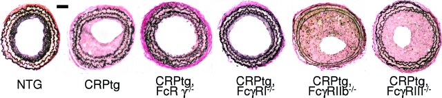
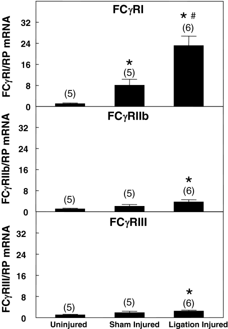
Neointima formation after vascular injury is exaggerated in ovariectomized (OVX) human C-reactive protein transgenic mice (CRPtg) compared to nontransgenic mice (NTG). We tested the hypothesis that this CRP-mediated exacerbation requires IgG Fc receptors (Fc gamma Rs). OVX NTG, CRPtg, and CRPtg lacking Fc gamma RI, Fc gamma RIIb, Fc gamma RIII, or the common gamma chain (FcR gamma) had their common carotid artery ligated. Twenty-eight days later neointimal thickening in CRPtg/Fc gamma RI(-/-) and CRPtg/FcR gamma (-/-) was significantly less than in CRPtg and no worse than in NTG, whereas in CRPtg/Fc gamma RIIb(-/-) and CRPtg/Fc gamma RIII(-/-) neointimal thickness was equal to or greater than in CRPtg. Immunohistochemistry revealed human CRP in the neointima of CRPtg, but little or none was observed in those lacking Fc gamma RI or FcR gamma. Real-time reverse transcriptase-polymerase chain reaction demonstrated that Fc gamma R types I to III were expressed in the CRPtg arteries, with Fc gamma RI expression increasing by threefold after ligation injury. Levels of serum complement (C3), neointimal deposition of complement (C3d), and cellular composition (monocytes, macrophages, lymphocytes) in the neointima did not differ among the different CRPtg genotypes. However, by immunofluorescence a neointimal population of F4/80+CRP+ cells was revealed only in OVX CRPtg. The exaggerated response to vascular injury provoked by CRP in OVX CRPtg depends on Fc gamma RI and probably requires its expression by F4/80+ cells.


Figures

Figure 1
Figure 1
Representative light micrographs of Verhoeff’s elastin-stained sections of carotid arteries harvested 28 days after ligation injury from OVX wild-type (NTG), CRPtg, and CRPtg FcγR-deficient mice. Note the narrow neointima in vessels from NTG, CRPtg/FcRγ−/−, and CRPtg/FcγRI−/− mice. Scale bar = 50 μm. Original magnifications, ×20.
Figure 2
Figure 2
Cross-sectional areas of neointima (A), media (B), and the resultant neointima/media ratio (C) of carotid arteries harvested 28 days after ligation injury from OVX wild-type (NTG), CRPtg, and CRPtg FcγR-deficient mice. Note that neointima area in vessels from CRPtg/FcRγ−/− and CRPtg/FcγRI−/− is comparable to that in NTG. Data shown are mean + SEM. Sample size (number of mice) is indicated. *P < 0.05 versus CRPtg.
Figure 3
Figure 3
Immunoreactive human CRP in carotid arteries 28 days after carotid artery ligation injury. Human CRP (brown-red staining) is evident in injured arteries of CRPtg (A), CRPtg/FcγRIIb−/− (D), and CRPtg/FcγRIII−/− (E) but little or no human CRP is detected in CRPtg/FcRγ−/− (B) and CRPtg/FcγRI−/− (C). Original magnifications, ×20.
Figure 4
Figure 4
FcγR expression (RT-PCR) in carotid arteries of OVX CRPtg. FcγRI was up-regulated after ligation injury compared to sham injury. Data are standardized to mean mRNA levels in uninjured arteries and are expressed as the mean ± SEM. *P < 0.05 versus uninjured; #P < 0.05 versus sham injured.
Figure 5
Figure 5
Complement deposition in ligation-injured carotid arteries. Despite widely variable neointima thickness, deposition of immunoreactive C3d (brown-red staining) is evident in injured arteries from all CRPtg genotypes. Original magnifications, ×20.
Figure 6
Figure 6
Cell types and their distribution in ligation-injured carotid arteries. Arteries were harvested from CRPtg (top) versus CRPtg/FcγRI−/− mice (bottom) 28 days after ligation injury, processed, and stained for immunoperoxidase-based detection of mouse F4/80 (monocytes/macrophages), B220 (B-cells), and CD4/CD8 (T cells). Mostly circulating monocytes and lymphocytes (punctuate staining) are seen in the patent vessels of CRPtg/FcγRI−/−, whereas in the more occluded vessels of CRPtg, these were fewer in number, and there were more cells within the proliferative area (stellate/diffuse staining). Importantly all three cell types were found in both genotypes. Original magnifications, ×20.
Figure 7
Figure 7
Human CRP is detected on monocytes/macrophages in the neointima of ligation-injured carotid arteries. Arteries were harvested from CRPtg mice 28 days after ligation injury, processed, and stained for immunofluorescence-based detection of human CRP, mouse F4/80, and mouse smooth muscle actin. The elastic laminae, which autofluoresce in the green channel, were used as a landmark. Left: Tissue sections were stained for human CRP (green) and mouse F4/80 (red); co-localization of CRP and F4/80 signals appears yellow-orange in the merged image (third panel from top). In the merged image the arrow points to a cell that stains for CRP only, whereas the box surrounds several F4/80-positive cells that also stain positive for human CRP. The area within the box is rotated and magnified and presented at the bottom of the left column. Right: Tissue sections were stained for human CRP (green) and mouse smooth muscle actin (red). Note that there is no evidence of co-localization in the merged image (third panel from the top). In the merged image the box surrounds a CRP-positive stellate cell, which is magnified and reproduced in the bottom panel. Original magnifications, ×20.

References

    1. van Leeuwen MA, van Rijswijk MH. Acute phase proteins in the monitoring of inflammatory disorders. Baillieres Clin Rheumatol. 1994;8:531–552. - PubMed
    1. Bassuk SS, Rifai N, Ridker PM. High-sensitivity C-reactive protein: clinical importance. Curr Probl Cardiol. 2004;29:439–493. - PubMed
    1. Pasceri V, Cheng JS, Willerson JT, Yeh ET. Modulation of C-reactive protein-mediated monocyte chemoattractant protein-1 induction in human endothelial cells by anti-atherosclerosis drugs. Circulation. 2001;103:2531–2534. - PubMed
    1. Pasceri V, Willerson JT, Yeh ET. Direct proinflammatory effect of C-reactive protein on human endothelial cells. Circulation. 2000;102:2165–2168. - PubMed
    1. Venugopal SK, Devaraj S, Yuhanna I, Shaul P, Jialal I. Demonstration that C-reactive protein decreases eNOS expression and bioactivity in human aortic endothelial cells. Circulation. 2002;106:1439–1441. - PubMed

Publication types