Review
. 2007 Dec;71(4):576-99.
doi: 10.1128/MMBR.00015-07.

Function, structure, and evolution of the RubisCO-like proteins and their RubisCO homologs

Affiliations
Review

Function, structure, and evolution of the RubisCO-like proteins and their RubisCO homologs

F Robert Tabita et al. Microbiol Mol Biol Rev. 2007 Dec.

Abstract

About 30 years have now passed since it was discovered that microbes synthesize RubisCO molecules that differ from the typical plant paradigm. RubisCOs of forms I, II, and III catalyze CO(2) fixation reactions, albeit for potentially different physiological purposes, while the RubisCO-like protein (RLP) (form IV RubisCO) has evolved, thus far at least, to catalyze reactions that are important for sulfur metabolism. RubisCO is the major global CO(2) fixation catalyst, and RLP is a somewhat related protein, exemplified by the fact that some of the latter proteins, along with RubisCO, catalyze similar enolization reactions as a part of their respective catalytic mechanisms. RLP in some organisms catalyzes a key reaction of a methionine salvage pathway, while in green sulfur bacteria, RLP plays a role in oxidative thiosulfate metabolism. In many organisms, the function of RLP is unknown. Indeed, there now appear to be at least six different clades of RLP molecules found in nature. Consideration of the many RubisCO (forms I, II, and III) and RLP (form IV) sequences in the database has subsequently led to a coherent picture of how these proteins may have evolved, with a form III RubisCO arising from the Methanomicrobia as the most likely ultimate source of all RubisCO and RLP lineages. In addition, structure-function analyses of RLP and RubisCO have provided information as to how the active sites of these proteins have evolved for their specific functions.


Figures

FIG. 1.
FIG. 1.
Unrooted NJ tree of RubisCO/RLP lineages. To construct this tree, a total of 193 sequences were aligned with MEGA 3.1 (38) and evaluated by ProtTest (1), and the tree was then constructed using the equal-input model with a gamma rate distribution of 1.554. The total numbers of sequences considered in each lineage were 35 for I-A, 16 for I-B, 9 for I-C, 22 for I-D, 20 for II, 10 for III-1, 4 for III-2, 20 for IV-NonPhoto, 2 for IV-EnvOnly, 14 for IV-Photo, 16 for IV-DeepYkrW, 12 for IV-YkrW, and 5 for IV-GOS. The width of the arrows is directly proportional to the number of sequences considered for each clade. For a complete list of sequences and sources, see Table S1 in the supplemental material. The scale bar represents a difference of 0.5 substitutions per site. Bootstrap values for nodes are shown in Fig. 2A. Single-sequence abbreviations and sequence identifiers are as follows: IV-Arc.ful-DSM 4304, Archaeoglobus fulgidus strain DSM4304 (GenBank accession number NP_070416); Met.bur-DSM6242, Methanococcoides burtonii strain DSM6242 (accession number ZP_00563653); Met.hun-JF-1, Methanospirillum hungatei strain JF-1 (accession number YP_503739); Met.the-PT, Methanosaeta thermophila strain PT (accession number ZP_01153096).
FIG. 2.
FIG. 2.
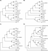
Comparison of RubisCO/RLP tree topologies reconstructed with NJ (A), ME (B), UPGMA (C), and MP (D). All except MP assumed a distribution of 1.554 of evolutionary rates across four categories as calculated by ProtTest (1). Values at nodes represent bootstrap support observed in 1,000 trials per method. IV-Arc.ful-DSM 4304, Archaeoglobus fulgidus strain DSM4304 (GenBank accession number NP_070416); Met.bur-DSM6242, Methanococcoides burtonii strain DSM6242 (accession number ZP_00563653); Met.hun-JF-1, Methanospirillum hungatei strain JF-1 (accession number YP_503739); Met.the-PT, Methanosaeta thermophila strain PT (accession number ZP_01153096).
FIG. 3.
FIG. 3.
Conservation of RubisCO active-site residues in RubisCO/RLP family members as noted previously by Cleland et al. (13) and Tabita (68). All form III RubisCO and RLP (form IV) sequences used in the reconstruction of phylogenetic relationships are included. Residues are noted in single-letter IUPAC code. Positions shaded green indicate conservation, while yellow indicates a semiconservative substitution and red indicates a nonconservative substitution. C, catalytic residue; R, RuBP binding residue.
FIG. 4.
FIG. 4.
Growth of four purple nonsulfur bacteria on MTA as the sole sulfur source. Rr, Rhodospirillum rubrum; Rp, Rhodopseudomonas palustris; Rc, Rhodobacter capsulatus; Rs, Rhodobacter sphaeroides. Growth on MTA correlates with the presence of RLP, further shown by inactivating the RLP gene (Singh and Tabita, unpublished).
FIG. 5.
FIG. 5.
Local conservation near genes encoding form III RubisCO (A) or the RLP lineages IV-Photo (B), IV-NonPhoto (C), and IV-YkrW. Gene neighborhoods were visualized using tools at the Integrated Microbial Genomes website (http://img.jgi.doe.gov/cgi-bin/pub/main.cgi). RubisCO/RLP genes are indicated in red. Other open reading frames are colored and identified according to their annotation in the Integrated Microbial Genomes database. Methyl-coM, methyl coenzyme M; Bchl, bacteriochlorophyll; Me-T'ferase, methyltransferase; 5-Me-C RE, 5-methyl-cytosine removing enzyme; EF-Ts, elongation factor Ts; SDR, short chain dehydrogenase/reductase.
FIG. 6.
FIG. 6.
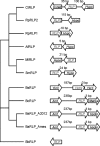
RLPs grouped by their functional linkage patterns. The 11 RLPs indicated here can be divided into two major groups. In the first group, all RLPs are linked to a hypothetical protein by the gene cluster method with short intergenic distances. The two hypothetical proteins next to the RLPs in Mesorhizobium loti and Sinorhizobium meliloti are homologous to each other. All the RLPs from Bacillus species form the second group. They have very similar gene organizations on the chromosome. They all reside between an aminotransferase and a hydrolase, which overlaps with RLPs by 3 bp. The RLP from Bordetella bronchiseptica does not have any functional linkages with high confidence. Oxred, oxidoreductase; Hypo, hypothetical protein; Amt, aminotransferase; MetSal, methylthioribose salvage protein; Hydro, hydrolase, haloacid dehalogenase-like hydrolase; CtRLP, C. tepidum RLP; RpRLP2, R. palustris RLP2; AfRLP A. fulgidus RLP; SmRLP, Sinorhizobium meliloti RLP; BsRLP, B. subtilis RLP; BcRLP, B. cereus RLP; BaRLP, B. anthracis RLP; BbRLP, Bordetella bronchiseptica RLP.
FIG. 7.
FIG. 7.
Methionine salvage pathway in which the YkrW-type RLP, such as the protein from B. subtilis (8), encoded by the mtnW/ykrW gene, participates in an enolase reaction whereby 2,3-diketo-5-methylthiopentyl-1-phosphate is converted to 2-hydroxy-3-keto-5-methylthiopentenyl-1-phosphate (highlighted). The products of the mtnX/ykrX, mtnZ/ykrZ, and mtnV/ykrV genes then allow methionine to be formed. SAM, S-adenosylmethionine. (Adapted from reference with permission of the publisher.)
FIG. 8.
FIG. 8.
RubisCO and B. subtilis RLP catalyze similar enolase-type reactions and employ structurally analogous substrates (see reference 33). In each instance, a carbamylated lysine catalyzes proton abstraction from the substrate to initialize enolization. DK-MTP 1-P, 2,3-diketo-5-methylthiopentyl-1-phosphate; HK-MTP 1-P, 2-hydroxy-3-keto-5-methylthiopentenyl-1-phosphate. (Adapted with permission from reference . Copyright 2007 American Chemical Society.)
FIG. 9.
FIG. 9.
Model for the evolution of RubisCO large subunits and RLP. The ancestor of all extant RubisCO large subunits and RLPs is proposed to have arisen in the Methanomicrobia with subsequent distribution by vertical transmission (solid arrows) and lateral transfer (dashed arrows) within the archaea. A central event in the evolutionary history was the acquisition of both a form III RubisCO and an RLP (IV-DeepYkr) by an ancestral eubacterium from the archaea. From these two ancestral sequences, diverse form I, form II, and form IV enzymes evolved within the Proteobacteria and Cyanobacteria and have been subsequently distributed by lateral gene transfer and by endosymbiotic events (dashed and dotted arrows) involving both Cyanobacteria and Alphaproteobacteria, leading to the phylogenetic distribution of sequences seen in nature today. The small subunit of form I RubisCO must have originated soon after the transfer of form III to the eubacterial ancestor prior to the divergence of Proteobacteria and Cyanobacteria.
FIG. 10.
FIG. 10.
The active sites in the crystal structures of form I (spinach) RubisCO (PDB accession number 8RUC) with bound CABP, C. tepidum RLP (PDB accession number 1YKW), R. palustris RLP2 (PDB accession number 2QYG), and G. kaustophilus RLP (accession number 2OEM) with bound DK-H-1-P. The side chains of active-site residues are shown as sticks, except for residue R383 in C. tepidum RLP and R. palustris RLP2. Only the backbone carbon and nitrogen atoms of R383 in the RLPs are shown. CABP and DK-H-1-P are shown in white, and the P1 and P2 phosphate groups are labeled in red and orange. Residues involved in contributing hydrogen bonds with the P1 phosphate group are green, residues involved in making hydrogen bonds with the backbone of CABP are orange, residues coordinating the Mg2+ atom (shown in magenta) are light red, and residues involved in binding P2 phosphate group are cyan. Not all parts of the structures are shown for the purpose of clarity.
FIG. 11.
FIG. 11.
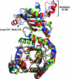
The monomer structures of RLP2 from R. palustris and RLP from G. kaustophilus superimposed with the RLP from C. tepidum. R. palustris RLP2 is blue, G. kaustophilus RLP is red, and C. tepidum RLP is green. The root mean square deviation (RMSD) of the Cα atom is 0.8 Å between R. palustris RLP2 and C. tepidum RLP, 1.3 Å between R. palustris RLP2 and G. kaustophilus RLP, and 1.3 Å between C. tepidum RLP2 and G. kaustophilus RLP. Two main structural differences can be seen in the N-terminal domain: loop CD in C. tepidum RLP and R. palustris RLP2 becomes a helix in G. kaustophilus RLP, and residues 47 to 58, missed in C. tepidum RLP, become a loop in R. palustris RLP2 and partly a helix in G. kaustophilus RLP.
FIG. 12.
FIG. 12.
Comparison of secondary structural elements in the X-ray crystal structures of the different forms of RubisCO. Large subunits from the structures of spinach (form I; PDB accession number 8RUC) (yellow), T. kodakarensis (form III; accession number 1GEH) (purple), and R. rubrum (form II; accession number 5RUB) (red) were superimposed on C. tepidum RLP (form IV; accession number 1YKW) (green) to align the α-carbon backbones. The transition state analog CABP (black sticks), which is present only in the spinach structure, has been drawn into the other structures to indicate the positions of active sites. A basic unit common to all four types of structures is formed as a result of the association of at least two of the large subunits. The active sites in bona fide RubisCO enzymes are contributed by residues from the N-terminal domain of one large subunit and the C-terminal domain of the other. Loop CD, which is present only in the RLPs and the RubisCO β-hairpin structure that is absent in the RLP structure, is indicated.
FIG. 13.
FIG. 13.
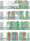
Structural alignment of representative sequences from RLPs and RubisCO large subunits. Superimposition of the X-ray crystal structures of C. tepidum RLP (PDB accession number 1YKW; form IV), spinach RubisCO (accession number 8RUC; form I), T. kodakarensis RubisCO (accession number 1GEH; form III), and R. rubrum RubisCO (accession number 5RUB; form II) was used to deduce the alignment of secondary structural elements (helices as bars and β-strands as arrows). Residue numbers are indicated on each side of the sequences. Conserved active-site residues are marked with an “*” below the sequences. RubisCO large-subunit sequences are boxed in gray. Residues that are identical or similar to those in other species are colored uniquely based on the nature of the residue. The catalytic loop 6, β-hairpin (both present in RubisCO enzymes), and loop CD (present only in RLPs) are indicated. A. vinosum, Allochromatium vinosum; C. limicola, Chlorobium limicola; O. granulosus, Oceanicola granulosus; P. horikoshii, Pyrococcus horikoshii; T. denitrificans, Thiobacillus denitrificans.
FIG. 13.
FIG. 13.
Structural alignment of representative sequences from RLPs and RubisCO large subunits. Superimposition of the X-ray crystal structures of C. tepidum RLP (PDB accession number 1YKW; form IV), spinach RubisCO (accession number 8RUC; form I), T. kodakarensis RubisCO (accession number 1GEH; form III), and R. rubrum RubisCO (accession number 5RUB; form II) was used to deduce the alignment of secondary structural elements (helices as bars and β-strands as arrows). Residue numbers are indicated on each side of the sequences. Conserved active-site residues are marked with an “*” below the sequences. RubisCO large-subunit sequences are boxed in gray. Residues that are identical or similar to those in other species are colored uniquely based on the nature of the residue. The catalytic loop 6, β-hairpin (both present in RubisCO enzymes), and loop CD (present only in RLPs) are indicated. A. vinosum, Allochromatium vinosum; C. limicola, Chlorobium limicola; O. granulosus, Oceanicola granulosus; P. horikoshii, Pyrococcus horikoshii; T. denitrificans, Thiobacillus denitrificans.
FIG. 14.
FIG. 14.
Comparison of the unique loop CD of C. tepidum RLP (PDB accession number 1YKW) (A) with the comparable region of form I (spinach) RubisCO (accession number 8RUC) (B). Residues Q78 to I91 form a loop (loop CD) (red ribbon and sticks), and residues in this loop have multiple interactions with residues of the same subunit (green ribbon and sticks) or the neighboring large subunit (purple ribbon and sticks). Notably, the hydroxyl group of S86 forms a hydrogen bond with loop 6 residue R327 (orange sticks) from the neighboring large subunit. Spinach form I residues equivalent to E75, E77, and H92 of C. tepidum RLP are E93, E94, and N95 (red ribbon and sticks). Residues K305 and V475 (yellow sticks) interact with E93 in the closed conformation of spinach RubisCO.
FIG. 15.
FIG. 15.
Placement of the β-hairpin residues in the holoenzyme structure of form I (spinach) RubisCO (PDB accession number 8RUC). The β-hairpin residues (Y353 to S367) (red) that are absent in RLPs are exposed to the solvent in the holoenzyme structure of spinach RubisCO. The large subunits are yellow, and the small subunits are blue.

References

    1. Abascal, F., R. Zardoya, and D. Posada. 2005. Prottest: selection of best-fit models of protein evolution. Bioinformatics 21:2104-2105. - PubMed
    1. Alfreider, A., C. Vogt, D. Hoffmann, and W. Babel. 2003. Diversity of ribulose-1,5-bisphosphate carboxylase/oxygenase large-subunit genes from groundwater and aquifer microorganisms. Microb. Ecol. 45:317-328. - PubMed
    1. Anantharaman, V., L. Aravind, and E. V. Koonin. 2003. Emergence of diverse biochemical activities in evolutionarily conserved structural scaffolds of proteins. Curr. Opin. Chem. Biol. 7:12-20. - PubMed
    1. Andersson, I. 1996. Large structures at high resolution: the 1.6 Å crystal structure of spinach ribulose-1,5-bisphosphate carboxylase/oxygenase complexed with 2-carboxyarabinitol bisphosphate. J. Mol. Biol. 259:160-174. - PubMed
    1. Andersson, I., and T. C. Taylor. 2003. Structural framework for catalysis and regulation in ribulose-1,5-bisphosphate carboxylase/oxygenase. Arch. Biochem. Biophys. 414:130-140. - PubMed

Publication types

MeSH terms

LinkOut - more resources