Review
. 2007 Dec;71(4):600-19.
doi: 10.1128/MMBR.00011-07.

The autodisplay story, from discovery to biotechnical and biomedical applications

Affiliations
Review

The autodisplay story, from discovery to biotechnical and biomedical applications

Joachim Jose et al. Microbiol Mol Biol Rev. 2007 Dec.

Abstract

Among the pathways used by gram-negative bacteria for protein secretion, the autotransporter pathway represents a solution of impressive simplicity. Proteins are transported, independent of their nature as recombinant or native passengers, as long as the coding nucleotide sequence is inserted in frame between those of an N-terminal signal peptide and a C-terminal domain, referred to as the beta-barrel of the outer membrane translocation unit. The immunoglobulin A1 (IgA1) protease from Neisseria gonorrhoeae was the first identified member of the autotransporter family of secreted proteins. The IgA1 protease was employed in initial experiments investigating autotransporter-mediated surface display of recombinant proteins and to investigate structural and functional requirements. Various other autotransporter proteins have since been described, and the autodisplay system was developed on the basis of the natural Escherichia coli autotransporter protein AIDA-I (adhesin involved in diffuse adherence). Autodisplay has been used for the surface display of random peptide libraries to successfully screen for novel enzyme inhibitors. The autodisplay system was also used for the surface display of functional enzymes, including esterases, oxidoreductases, and electron transfer proteins. Whole E. coli cells displaying enzymes have been utilized to efficiently synthesize industrially important rare organic compounds with specific chirality. Autodisplay of epitopes on the surface of attenuated Salmonella carriers has also provided a novel way to induce immune protection after oral vaccination. This review summarizes the structural and functional features of the autodisplay system, illustrating its discovery and most recent applications. Autodisplay facilitates the export of more than 100,000 recombinant molecules per single cell and permits the oligomerization of subunits on the cell surface as well as the incorporation of inorganic prosthetic groups after transport of apoproteins onto the bacterial surface without disturbing bacterial integrity or viability. We discuss future biotechnical and biomedical applications in the light of these achievements.


Figures

FIG. 1.
FIG. 1.
Secretion mechanism of the autotransporter proteins. (A) Structure of the polyprotein precursor. (B) Transport of the recombinant passenger. By the use of a typical signal peptide, a precursor protein is transported across the inner membrane. After arrival at the periplasm, the C-terminal part of the precursor folds as a porin-like structure, a so-called β-barrel within the outer membrane, and the passenger is transmitted to the cell surface. SP, signal peptide; IM, inner membrane; PP, periplasm; OM, outer membrane.
FIG. 2.
FIG. 2.
Linear structure of the CTB-IgAβ hybrid protein, used for the first surface display of a recombinant protein with the aid of an autotransporter. SP, signal peptide; P, passenger.
FIG. 3.
FIG. 3.
Structure of an artificial autotransporter protein as used in autodisplay. FP-CT is a cysteine-containing fusion protein that is encoded either by plasmid pSH4 under control of a strong constitutive promoter (PTK) or by plasmid pET-SH4 under the control of the inducible T7/lac promoter. The environments of the passenger insertion site, necessary to obtain surface translocation, are given as sequences. Restriction endonuclease cleavage sites for insertion of passenger-encoding DNA sequences are underlined. Various other restriction sites are available in similar plasmids. The signal peptide originates from CTB, and the signal peptidase cleavage site is marked by an arrow. The signal peptides of PelB, AIDA-I, OmpA, and β-lactamase have also been used in combination with translocation units from different natural autotransporter proteins. The specific cleavage site for IgA1 protease that can be applied for the release of the passenger protein into the extracellular milieu is in italics, and the linear epitope for a mouse monoclonal antibody (Dü142) which can be used for labeling is in bold. The cysteine used in “cystope tagging,” a specific labeling and detection method, is indicated by a black box.
FIG. 4.
FIG. 4.
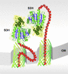
Passenger-driven dimerization of SDH expressed by autodisplay at the cell surface. Due to the free motility of the β-barrel, serving as an anchor within the outer membrane in autodisplay, passenger proteins can spontaneously form dimers at the cell surface, even when they are expressed as monomers form monomeric genes. This is aided by the high number of recombinant proteins (e.g., SDH) expressed at the cell surface by autodisplay, bringing the monomers in close enough vicinity to interact. It is a unique feature of the autodisplay system and has not been reported for any other surface display system so far.
FIG. 5.
FIG. 5.
Whole-cell biocatalyst for the synthesis of steroids obtained by autodisplay of Adx. After transport of apo-Adx to the cell surface, dimers were formed spontaneously and the electron transfer activity of Adx was restored by chemical incorporation of the [2Fe-2S] cluster. After the addition of purified AdR and CYP11A1 or CYP11B1, an efficient whole-cell biocatalyst for the synthesis of pregnenolone or corticosterone, respectively, was obtained. The cell envelope of E. coli provided a membrane environment sufficient for both P450 enzymes to be active. OM, outer membrane.
FIG. 6.
FIG. 6.
Distribution of the β-barrels within the outer membrane of E. coli by the use of the autodisplay system. (A) The rod-shaped cell of E. coli can be idealized as a cylinder with a height of 5 μm and a radius of 0.5 μm. with these values, the entire surface area of a single cell can be calculated to be 17.2 μm2. (B) The number of active Adx molecules displayed at the surface of a single cell using autodisplay has experimentally been determined to be 180,000. The radius of a single β-barrel can be calculated to be 1.1 nm, which is in congruence with the value derived from the crystal structure of NalP (1.0 nm in one dimension and 1.25 nm in the other). Given that the barrels are evenly spread over the entire surface, the mean distance between two adjacent molecules can be calculated to be 8.4 nm in any dimension by assuming that the expansion space of one barrel has the shape of an equilateral hexagon. (C) E. coli cells displaying 180,000 Adx molecules were subjected to indirect immunofluorescence microscopy. After labeling with an anti-Adx rabbit serum and a second anti-rabbit antibody coupled to FITC, cells were analyzed, and no indication was found for a polar distribution of recombinant Adx molecules on the cell surface of E. coli as has been reported for the natural passenger of AIDA-I. Rather, the picture obtained accords with an even distribution all over the entire surface.
FIG. 7.
FIG. 7.
Surface display library screening to identify new enzyme inhibitors by autodisplay. (1) A peptide that is known to be an inhibitor (the lead) is expressed at the cell surface by autodisplay and tested for functionality. (2) Random libraries are generated by standard genetic engineering tools, where each cell of E. coli displays one distinct variant in high numbers. (3) The library of E. coli cells with different variants is screened by target enzyme labeling. Since an enzyme inhibitor has high affinity to its target enzyme and as a consequence the enzyme has high affinity to the inhibitor as well, the target enzyme will bind to an inhibiting structure expressed at the cell surface. If the target enzyme is coupled to a fluorescent dye (e.g., FITC), the cell displaying the appropriate variant can be sorted by FACS. (4) The cell selected by this procedure can be used for clonal production of cell quantities sufficient for, e.g., DNA sequence analysis and hence for the identification of a new lead structure.

References

    1. Adams, T. M., A. Wentzel, and H. Kolmar. 2005. Intimin-mediated export of passenger proteins requires maintenance of a translocation-competent conformation. J. Bacteriol. 187:522-533. - PMC - PubMed
    1. Barbar, E., G. Barany, and C. Woodward. 1996. Unfolded BPTI variants with a single disulfide bond have diminished non-native structure distant from the crosslink. Fold. Des. 1:65-76. - PubMed
    1. Becker, S., H. U. Schmoldt, T. M. Adams, S. Wilhelm, and H. Kolmar. 2004. Ultra-high-throughput screening based on cell-surface display and fluorescence-activated cell sorting for the identification of novel biocatalysts. Curr. Opin. Biotechnol. 15:323-329. - PubMed
    1. Becker, S., S. Theile, N. Heppeler, A. Michalczyk, A. Wentzel, S. Wilhelm, K. E. Jaeger, and H. Kolmar. 2005. A generic system for the Escherichia coli cell-surface display of lipolytic enzymes. FEBS Lett. 579:1177-1182. - PubMed
    1. Benhar, I. 2001. Biotechnological applications of phage and cell display. Biotechnol. Adv. 19:1-33. - PubMed

MeSH terms

LinkOut - more resources