Editorial
. 2007 Dec 7;101(12):1213-5.
doi: 10.1161/CIRCRESAHA.107.165662.

The carboxy terminal domain of connexin43: from molecular regulation of the gap junction channel to supramolecular organization of the intercalated disk

Editorial

The carboxy terminal domain of connexin43: from molecular regulation of the gap junction channel to supramolecular organization of the intercalated disk

Nicholas J Severs. Circ Res. .
No abstract available


Figures

None
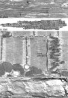
Supramolecular organization of junctions at the intercalated disk. Light microscopy of ventricular myocardium (A) shows the disks as dense irregular dark lines (arrows), marking the boundaries between individual myocytes. If we take a single myocyte (between squared brackets), and view it at higher magnification by thin-section electron microscopy (B), the step-like features of the disks are clearly seen (right end of cell and arrowheads). If we then take, at higher magnification, an area from within the disk like that enclosed by the box in (B), the three junction types are visible (C). Fasciae adherentes occupy electron-dense vertical plicate zones of the disk; gap junctions and desmosomes mainly the lateral-facing zones. Note that each end of the gap junction in this example touches a fascia adherens (arrowheads). However, not all gap junctions make such direct contact with adhesion junctions; the longitudinal zone of disk membrane shown by freeze-fracture electron microscopy in (D) extends over three sarcomeres, with three gap junctions (areas outlined with dashes). To the right (between arrowheads), the membrane turns up at right angles toward the viewer into a fascia adherens-containing plicate zone. Each ventricular myocyte is linked to ∼10 other myocytes by intercalated disks. Although all disks are constructed according to a common plan, they vary considerably in size and morphology. The largest disk occupies the entire maximal width of the myocyte body whereas the smallest is formed from single abutting myofibrils (extending as side branches from the cell bodies). The disk may appear as a series of steps that change course at the position of consecutive sarcomeres of adjacent myofibrils; the longitudinal zones may extend for one, two, three or more sarcomeres in length. The steps may form a continuous series or they may go back and forth so that each end of the disk is in line. Within any given disk, there may be many steps or few. (B, from Severs et al with permission from Elsevier; C from Severs17).

Comment on

References

    1. Severs NJ. Review. The cardiac gap junction and intercalated disc. Int J Cardiol. 1990;26:137–173. - PubMed
    1. Gourdie RG, Green CR, Severs NJ. Gap junction distribution in adult mammalian myocardium revealed by an antipeptide antibody and laser scanning confocal microscopy. J Cell Sci. 1991;99:41–55. - PubMed
    1. Hoyt RH, Cohen ML, Saffitz JE. Distribution and three-dimensional structure of intercellular junctions in canine myocardium. Circ Res. 1989;64:563–574. - PubMed
    1. Severs NJ. Gap junction shape and orientation at the cardiac intercalated disk. Circ Res. 1989;65:1458–1461. - PubMed
    1. Severs NJ, Shovel KS, Slade AM, Powell T, Twist VW, Green CR. Fate of gap junctions in isolated adult mammalian cardiomyocytes. Circ Res. 1989;65:22–42. - PubMed