Comparative Study
. 2008 May;38(5):501-8.
doi: 10.1165/rcmb.2007-0298OC. Epub 2007 Dec 6.

Estrogen determines sex differences in airway responsiveness after allergen exposure

Affiliations
Comparative Study

Estrogen determines sex differences in airway responsiveness after allergen exposure

Shigeki Matsubara et al. Am J Respir Cell Mol Biol. 2008 May.

Abstract

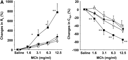
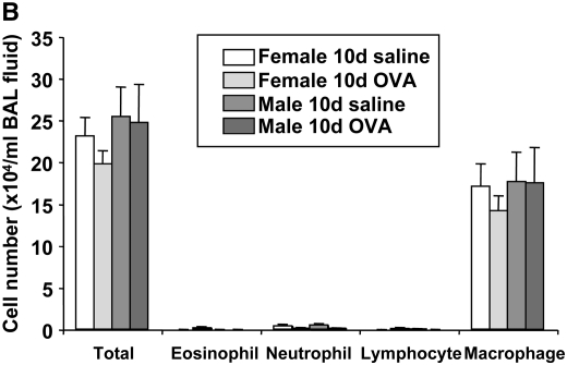
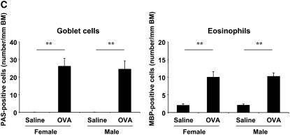
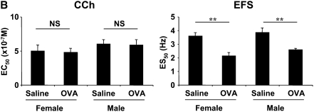
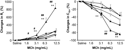
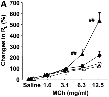
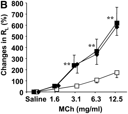
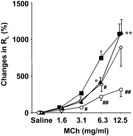
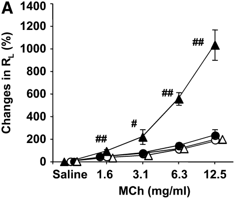
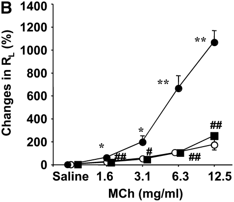
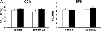
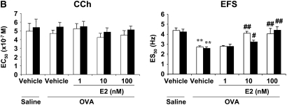
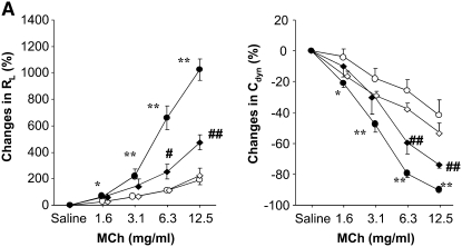
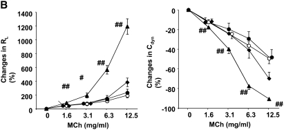
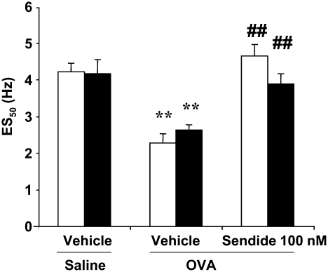
The female hormone estrogen is an important factor in the regulation of airway function and inflammation, and sex differences in the prevalence of asthma are well described. Using an animal model, we determined how sex differences may underlie the development of altered airway function in response to allergen exposure. We compared sex differences in the development of airway hyperresponsiveness (AHR) after allergen exposure exclusively via the airways. Ovalbumin (OVA) was administered by nebulization on 10 consecutive days in BALB/c mice. After methacholine challenge, significant AHR developed in male mice but not in female mice. Ovariectomized female mice showed significant AHR after 10-day OVA inhalation. ICI182,780, an estrogen antagonist, similarly enhanced airway responsiveness even when administered 1 hour before assay. In contrast, 17beta-estradiol dose-dependently suppressed AHR in male mice. In all cases, airway responsiveness was inhibited by the administration of a neurokinin 1 receptor antagonist. These results demonstrate that sex differences in 10-day OVA-induced AHR are due to endogenous estrogen, which negatively regulates airway responsiveness in female mice. Cumulatively, the results suggest that endogenous estrogen may regulate the neurokinin 1-dependent prejunctional activation of airway smooth muscle in allergen-exposed mice.


Figures

<b>Figure 1.</b>
Figure 1.
Comparison of airway responsiveness and inflammation in female and male mice after 10-day ovalbumin (OVA) inhalation. Female and male mice were exposed to either a 0.9% saline solution or a 1% OVA solution for 10 days (20 min/d). (A) Airway hyperresponsiveness (AHR) to inhaled methacholine (MCh) after 10-day OVA inhalation. AHR (airway resistance [RL] and lung dynamic compliance [Cdyn]) to inhaled MCh was assayed 24 hours after the last OVA challenge. Circles, female mice; squares, male mice; open symbols, 10-day saline; solid symbols, 10-day OVA. (B) Bronchoalveolar lavage (BAL) cell composition after 10-day OVA inhalation. BAL fluid was recovered immediately after the AHR assay was completed. (C) Numbers of periodic acid-Schiff–positive and major basic protein–positive cells. Results represent the mean ± SEM (n = 8). *P < 0.05 and **P < 0.01 versus 10-day saline exposure in each sex. ND = not detected. The baseline values of RL and Cdyn were as follows: 0.57 ± 0.03 cm H2O/ml/second (n = 16) and 0.069 ± 0.003 ml/cm H2O (n = 16) in female saline-treated mice, respectively; 0.59 ± 0.03 cm H2O/ml/s (n = 12) and 0.070 ± 0.002 ml/cm H2O (n = 12) in female OVA-treated mice, respectively; 0.55 ± 0.02 cm H2O/ml/second (n = 16) and 0.065 ± 0.003 ml/cm H2O (n = 16) in male saline-treated mice, respectively; 0.57 ± 0.02 cm H2O/ml/second (n = 21) and 0.064 ± 0.003 ml/cm H2O (n = 21) in male OVA-treated mice, respectively. BM = basement membrane.
<b>Figure 2.</b>
Figure 2.
Tracheal smooth muscle responsiveness to carbachol (CCh) and electrical field stimulation (EFS) after 10-day OVA inhalation. (A) Representative CCh and EFS responses. Top panels: Solid circles, female saline; open squares, female OVA. Bottom panels: Solid circles, male saline; open squares, male OVA. (B) Summary of results of CCh- and EFS-induced contractility. Results represent the mean ± SEM (n = 8). **P < 0.01 statistically significant. NS = not significant. 95% confidence intervals overlapped among the groups.
<b>Figure 3.</b>
Figure 3.
Development of AHR to inhaled MCh after 10-day OVA inhalation in ovariectomized (OVX) female mice. Sham-operated or OVX female mice were exposed to 0.9% saline or 1% OVA solution for 10 days. AHR was assayed 24 hours after the last OVA challenge. Sham operation or OVX was performed at least 2 weeks before initiation of allergen exposure. 17β-estradiol (E2) at a dose of 100 μg/kg was administered intraperitoneally to OVX mice 1 hour before assay of AHR. Results represent the mean ± SEM (n = 8). **P < 0.01 versus sham/OVA (open triangles) or OVX/saline (solid circles). #P < 0.05 and ##P < 0.01 versus sham/saline (open circles) or OVX/OVA/E2 (squares). Solid triangles, OVX/OVA.
<b>Figure 4.</b>
Figure 4.
Estrogen receptor antagonist alters AHR in female mice. (A) Effect of daily administration of an estrogen antagonist on AHR in female mice. Open circles, 10 days saline/10 days vehicle; solid circles, 10 days OVA/10 days vehicle; open triangles, 10 days saline/10 days ICI (10 mg/kg/d); solid triangles, 10 days OVA/10 days ICI (10 mg/kg/d). (B) Effect of daily administration of the antagonist in male mice after 10-day OVA inhalation in vivo. Open squares, 10 days saline/10 days vehicle; solid squares, 10 days OVA/10 days vehicle; diamonds, 10 days OVA/10 days ICI (10 mg/kg/d). AHR was assayed 24 hours after the last OVA challenge. ICI182,780 at a dose of 10 mg/kg/d was administered intraperitoneally immediately before each OVA challenge. Results represent the mean ± SEM (n = 8). **P < 0.01 and ##P < 0.01 comparing 10-day saline/10-day vehicle and 10-day OVA/10-day vehicle.
<b>Figure 5.</b>
Figure 5.
Effect of daily administration of E2 on 10-day OVA-induced AHR in male mice in vivo. AHR was assayed 24 hours after the last OVA challenge. E2 at doses of 50 and 100 μg/kg was administered intraperitoneally immediately before each OVA challenge. Results represent the mean ± SEM (n = 8). #P < 0.05 and ##P < 0.01 comparing 10-day OVA/10-day vehicle (squares); *P < 0.05 and **P < 0.01 comparing 10-day OVA/10-day E2 100 μg/kg/day (circles). Diamonds, 10-day OVA/10-day E2 (50 μg/kg/d); triangles, 10-day OVA/10-day E2 (μg/kg/d) + 10-day ICI.
<b>Figure 6.</b>
Figure 6.
Rapid effect of (A) an estrogen antagonist and (B) exogenous E2 on airway responsiveness to MCh in vivo. AHR was assayed 24 hours after the last OVA challenge. ICI182,780 (ICI) at a dose of 10 mg/kg to female mice (A) or E2 at a dose of 100 μg/kg to male mice (B) was administered intraperitoneally 1 hour before the AHR assay. Results represent the mean ± SEM (n = 8). #P < 0.05 and ##P < 0.01 comparing 10-day OVA/vehicle. *P < 0.05 and **P < 0.01 comparing 10-day saline/vehicle. Open circles, 10-day saline/vehicle; solid circles, 10-day OVA/vehicle; open triangles, 10-day saline/ICI; solid triangles, 10-day OVA ICI. Squares, 10-day OVA/E2.
<b>Figure 7.</b>
Figure 7.
Effect of exogenous E2 on in vitro airway responsiveness to CCh or EFS after (A) 10-day saline or (B) 10-day OVA exposure. Tracheas from female (open bars) and male (solid bars) mice were isolated from 10-day saline- or OVA-treated mice. The tracheas were preincubated with vehicle or E2 (1–100 nM) for 30 minutes. After incubation, airway contractility to CCh or EFS was determined. Results represent the mean ± SEM (n = 8). **P < 0.01 comparing 10-day saline/vehicle group in either sex. #P < 0.05 and ##P < 0.01 comparing 10-day OVA/vehicle group in either sex. In the CCh and EFS groups, the 95% confidence intervals overlapped.
<b>Figure 8.</b>
Figure 8.
Effect of a neurokinin-1 receptor antagonist (Sendide) on (A) 10-day OVA-induced AHR in male mice and (B) enhancement of AHR by an estrogen antagonist in 10-day OVA-exposed female mice. AHR was assayed 24 hours after the last OVA challenge. Sendide at a dose of 40 nmol/kg and/or ICI182,780 at a dose of 10 mg/kg was administered intraperitoneally 2 hours and/or 1 hour, respectively, before the AHR assay. Results represent the mean ± SEM (n = 8). #P < 0.05 and ##P < 0.01 comparing 10-day OVA/vehicle or 10-day saline/Sendide. *P < 0.05 and **P < 0.01 comparing 10-day saline/vehicle or 10-day saline/Sendide. P < 0.05 and ††P < 0.01 comparing 10-day OVA/ICI/PBS. PBS was a vehicle for Sendide. ICI = ICI182,780. A: open circles, 10-day saline/vehicle; solid circles, 10-day OVA/vehicle; open diamonds, 10-day saline/Sendide; solid diamonds, 10-day OVA/Sendide. B: open circles, 10-day saline/vehicle/PBS; solid circles, 10-day OVA/vehicle/PBS; triangles, 10-day OVA/ICI/PBS; diamonds, 10-day OVA/ICI/Sendide.
<b>Figure 9.</b>
Figure 9.
Involvement of neurokinin-1 in 10-day OVA-induced AHR detected by EFS in vitro. Tracheas from female (open bars) and male (solid bars) mice were isolated from 10-day saline- or OVA-exposed mice. The tracheas were preincubated with vehicle or Sendide (100 nM) for 30 minutes. After the preincubation, airway contractility to EFS was assayed. Results represent the mean ± SEM (n = 8). ††P < 0.01 comparing 10-day saline/vehicle. ##P < 0.01 comparing 10-day OVA/vehicle. ES50 = 50% of maximal contraction.

Comment in

Similar articles

Cited by

References

    1. Meyer MR, Haas E, Barton M. Sex differences of cardiovascular disease: new perspectives for estrogen receptor signaling. Hypertension 2006;47:1019–1026. - PubMed
    1. Most W, van der Wee-Pals L, Ederveen A, Papapoulos S, Lowik C. Ovariectomy and orchidectomy induce a transient increase in the osteoclastogenic potential of bone marrow cells in the mouse. Bone 1997;20:27–30. - PubMed
    1. Gillies GE, Murray HE, Dexter D, McArthur S. Sex dimorphisms in the neuroprotective effects of estrogen in an animal model of Parkinson's disease. Pharmacol Biochem Behav 2004;78:513–522. - PubMed
    1. Dimitropoulou C, White RE, Ownby DR, Catravas JD. Estrogen reduces carbachol-induced constriction of asthmatic airways by stimulating large-conductance voltage and calcium-dependent potassium channels. Am J Respir Cell Mol Biol 2005;32:239–247. - PubMed
    1. Degano B, Prevost MC, Berger P, Molimard M, Pontier S, Rami J, Escamilla R. Estradiol decreases the acetylcholine-elicited airway reactivity in ovariectomized rats through an increase in epithelial acetylcholinesterase activity. Am J Respir Crit Care Med 2001;164:1849–1854. - PubMed

Publication types

MeSH terms