

Evaluation of a new scoring system for retinal nerve fiber layer photography using HRA1 in 964 eyes
- PMID: 18063886
- PMCID: PMC2629886
- DOI: 10.3341/kjo.2007.21.4.216
Evaluation of a new scoring system for retinal nerve fiber layer photography using HRA1 in 964 eyes
Abstract
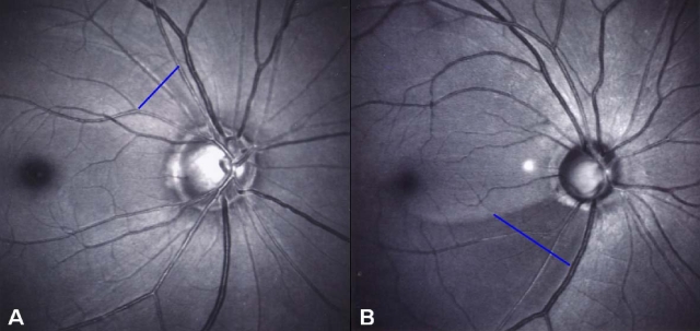
Purpose: To evaluate retinal nerve fiber layer (RNFL) defect by a new scoring system for RNFL photography using the Heidelberg Retina Angiograph 1 (HRA1).
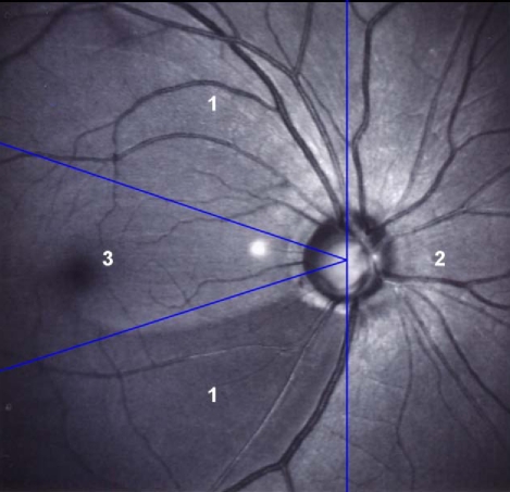
Methods: This retrospective study included 128 healthy eyes and 836 primary open-angle glaucoma eyes. The RNFL photography using HRA1 was interpreted using a new scoring system, and correlated with visual field indices of standard automated perimetry (SAP). Using the presence of RNFL defect, darkness, width, and location, we established the new scoring system of RNFL photos.
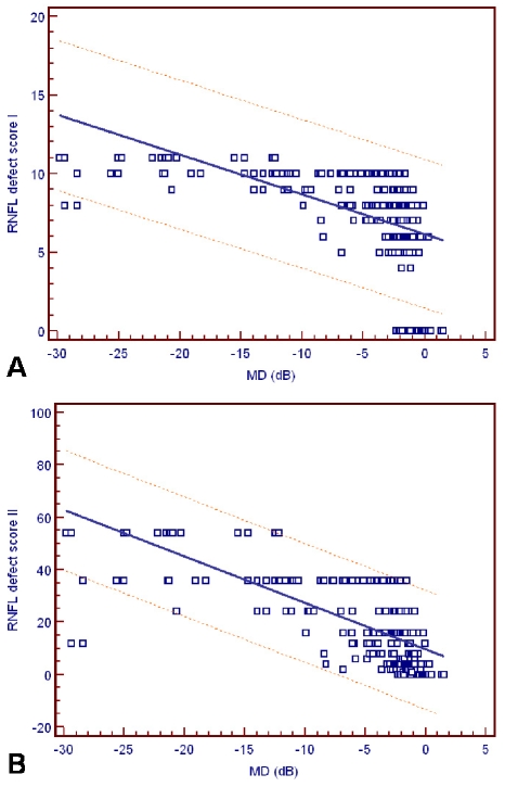
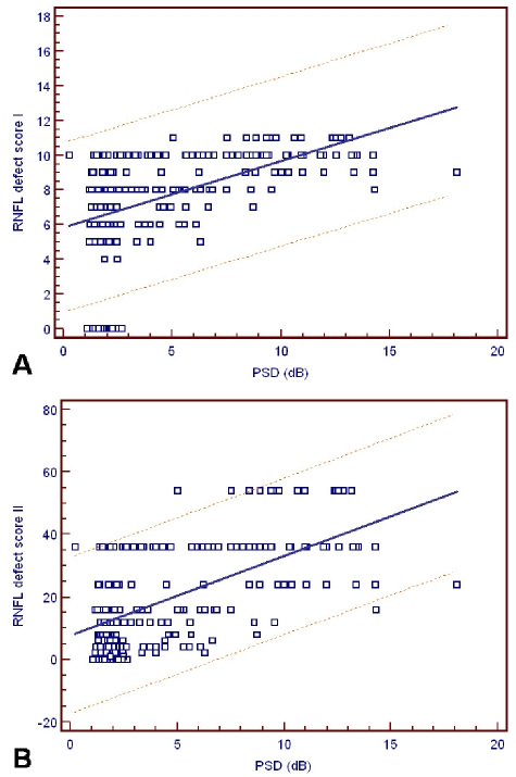
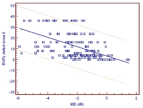
Results: The mean RNFL defect score I in the early, moderate, severe, and control groups were 7.3, 9.2, 10.4, and 3.6, respectively. The mean RNFL defect score II in the early, moderate, severe, and control groups were 14.5, 28.5, 43.4, and 3.4, respectively. Correlations between the RNFL defect score II and the mean deviation of SAP was the strongest of the various combinations (r=-0.675, P<.001).
Conclusions: Using a new scoring system, we propose a method for semi-quantitative interpretation of RNFL photographs. This scoring system may be helpful to distinguish between normal and glaucomatous eyes, and the score is associated with the severity of visual field loss.
Figures
Similar articles
-
Early glaucoma detection using the Humphrey Matrix Perimeter, GDx VCC, Stratus OCT, and retinal nerve fiber layer photography.Ophthalmology. 2007 Feb;114(2):210-5. doi: 10.1016/j.ophtha.2006.09.021. Ophthalmology. 2007. PMID: 17270671
-
Defective angles of localized retinal nerve fiber layer reflect the severity of visual field defect- a cross-sectional analysis.BMC Ophthalmol. 2020 Apr 9;20(1):141. doi: 10.1186/s12886-020-01396-y. BMC Ophthalmol. 2020. PMID: 32272929 Free PMC article.
-
A comparison of optical coherence tomography and retinal nerve fiber layer photography for detection of nerve fiber layer damage in glaucoma.Ophthalmology. 2000 Jul;107(7):1309-15. doi: 10.1016/s0161-6420(00)00168-8. Ophthalmology. 2000. PMID: 10889104 Clinical Trial.
-
Optic disc hemorrhage may be associated with retinal nerve fiber loss in otherwise normal eyes.Ophthalmology. 2008 Dec;115(12):2132-40. doi: 10.1016/j.ophtha.2008.08.024. Ophthalmology. 2008. PMID: 19041474
-
Diffuse glaucomatous structural and functional damage in the hemifield without significant pattern loss.Arch Ophthalmol. 2009 Nov;127(11):1442-8. doi: 10.1001/archophthalmol.2009.196. Arch Ophthalmol. 2009. PMID: 19901209
Cited by
-
Comparison of Blue and Green Confocal Scanning Laser Ophthalmoscope Imaging to Detect Retinal Nerve Fiber Layer Defects.Korean J Ophthalmol. 2019 Apr;33(2):131-137. doi: 10.3341/kjo.2018.0075. Korean J Ophthalmol. 2019. PMID: 30977322 Free PMC article.
-
Relationship between Peripapillary Retinal Nerve Fiber Layer Thickness Measured by Optical Coherence Tomography and Visual Field Severity Indices.Korean J Ophthalmol. 2015 Aug;29(4):263-9. doi: 10.3341/kjo.2015.29.4.263. Epub 2015 Jul 21. Korean J Ophthalmol. 2015. PMID: 26240511 Free PMC article.
References
-
- Hoyt WF, Frisen L, Newman NM. Fundoscopy of nerve fiber layer defects in glaucoma. Invest Ophthalmol. 1973;12:814–829. - PubMed
-
- Quigley HA, Katz J, Derick RJ, et al. An evaluation of optic disc and nerve fiber layer examinations in monitoring progression of early glaucoma damage. Ophthalmology. 1992;99:19–28. - PubMed
-
- Sommer A. Retinal nerve fiber layer. Am J Ophthalmol. 1995;120:665–667. - PubMed
-
- Sommer A, Katz J, Quigley HA, et al. Clinically detectable nerve fiber atrophy precedes the onset of glaucomatous field loss. Arch Ophthalmol. 1991;109:77–83. - PubMed
-
- Sommer A, Miller NR, Pollack I, et al. The nerve fiber layer in the diagnosis of glaucoma. Arch Ophthalmol. 1977;95:2149–2156. - PubMed
Publication types
MeSH terms
LinkOut - more resources
Full Text Sources
Miscellaneous
