Review
. 2008 Jan;7(1):68-83.
doi: 10.1038/nrd2463.

Neurophysiological biomarkers for drug development in schizophrenia

Affiliations
Review

Neurophysiological biomarkers for drug development in schizophrenia

Daniel C Javitt et al. Nat Rev Drug Discov. 2008 Jan.

Abstract

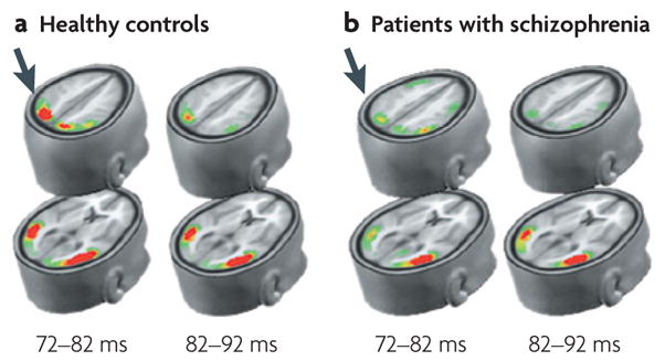
Schizophrenia represents a pervasive deficit in brain function, leading to hallucinations and delusions, social withdrawal and a decline in cognitive performance. As the underlying genetic and neuronal abnormalities in schizophrenia are largely unknown, it is challenging to measure the severity of its symptoms objectively, or to design and evaluate psychotherapeutic interventions. Recent advances in neurophysiological techniques provide new opportunities to measure abnormal brain functions in patients with schizophrenia and to compare these with drug-induced alterations. Moreover, many of these neurophysiological processes are phylogenetically conserved and can be modelled in preclinical studies, offering unique opportunities for use as translational biomarkers in schizophrenia drug discovery.


Figures

Figure 1
Figure 1. This figure reflects the multiple brain regions implicated in schizophrenia, and source of likely generators for specific biomarkers used in the study of schizophrenia
AX-CPT, AX-type visual continuous performance task; MMN, mismatch negativity; Ncl, closure negativity; SPEM, smooth pursuit eye movements.
Figure 2
Figure 2. Schematic diagram of mismatch negativity (MMN) generators in schizophrenia
a. MMN is elicited in an auditory oddball paradigm in which a sequence of repetitive standard stimuli (blue boxes) are interrupted by stimuli that differ in a physical stimulus dimension such as pitch or duration (green boxes). The deviant probability equals the number of deviants divided by the total number of stimuli. MMN reflects N-methyl-D-aspartate (NMDA)-dependent processing of stimulus deviance within the auditory sensory cortex. b. Schematic diagram of MMN generators within the auditory cortex (located in the superior temporal lobe, shown in red). Because of the orientation of MMN generators, the MMN reverses in polarity between the frontal midline electrode (Fz) and the left (LM) and right (RM) mastoids. Because pitch deviance can be detected at stimulus onset, but duration deviance can only be detected at the time of standard stimulus offset, duration MMN (pale blue line) is delayed relative to pitch (frequency) MMN (pink line). The dashed arrow indicates the orientation of the electrical field originating from the auditory cortex. Activity from auditory cortex characteristically inverts between the central midline electrode (Fz) and the mastoids (RM, LM), relative to a nose reference (not shown). c. Characteristic waveforms at Fz from patients with recent onset or chronic schizophrenia versus controls. Peak MMN responses (arrows) are significantly reduced in patients with schizophrenia relative to controls, for both pitch (top line) and duration (bottom line). Dashed lines illustrate the latency shift in response to pitch versus duration to deviant stimuli. Δf, pitch difference between standard and deviant; ERP, event-related potential; ISI, interstimulus interval. FIGS 2a and 2c are modified with permission from REF. © (2005) and REF. © (2006) Elsevier Science, respectively.
Figure 3
Figure 3. Visual P1 deficit in schizophrenia using local autoregressive average (LAURA) model
LAURA depicts the degree of brain electrical activity (current source density) within derived source regions. P1 reflects early stimulus-elicited activity within the dorsal (top rows) and ventral (bottom rows) visual streams, occurring within the first 100 ms following stimulation. In schizophrenia, activation is normal within the ventral stream, but is markedly reduced within the dorsal stream (arrows), reflecting impaired activity within the magnocellular system. This image is modified with permission from REF. © (2005) Oxford University Press.
Figure 4
Figure 4. Time/frequency domain electroencephalography measures from a control subject and a patient with chronic schizophrenia
The average event-related potential (ERP) (n = 88 trials) reveals that both P1 and N1 components are markedly reduced in the patient with schizophrenia compared with the control subject. Reduced activity across the frequency spectrum is apparent in the maps of evoked power, induced power and phase-locking synchrony (colour scales same as in BOX 1).
Figure 5
Figure 5. Cortical response variability is increased in patients with schizophrenia
a. Frequency domain analyses showing increased prefrontal noise, that is, increased variability of slow-wave oscillations, in patients with schizophrenia and their clinically unaffected siblings. Increased variability of slow-wave oscillations results from impaired phase-locking of these oscillations in schizophrenia. b. An analogous increase in variability of blood-oxygen-level dependent response is observed in patients with schizophrenia: group contrast analysis compared with controls. Prefrontal noise is modulated by synaptic dopamine signalling (catechol-O-methyltransferase (COMT)-genotype) as measured by electrophysiology and functional magnetic resonance imaging,,. This image is reproduced with permission from REF. © (2004) and REF. © (2006) American Psychiatric Association.
Figure 6
Figure 6. Components of the smooth pursuit eye movements: the underlying neural circuit
The smooth pursuit eye movement system functions to maintain the image of a moving object on the fovea by minimizing error between the target velocity and the eye velocity. Broadly, the system is divided into two components: the response based on the internal representation of the target motion (that is, the extraretinal motion information), and the corrections based on the visual feedback of discrepancies between the eye and target velocities, the so-called retinal error. The pursuit maintenance in a control subject (red graph) and a patient with schizophrenia (blue graph) is shown (a). The healthy individual is able to accurately match the target velocity, as this response is mostly driven by the extraretinal motion signals during the pursuit maintenance, whereas the patient with schizophrenia shows lower eye velocity, because of inadequate extraretinal motion signals that create a retinal error. In response to the visual feedback of the retinal error, the subject makes corrective responses in the form of a saccade or an increase in eye velocity (marked by Z in panel a). However, this increase in velocity is transient as the retinal error signal becomes weak as the eye velocity matches the target velocity. This interaction between the responses to retinal and extraretinal motion signals is modelled by the regression equation: maintenance of eye velocity = be × retinal error + ber × extraretinal error (where be and ber are coefficients associated with retinal error and extraretinal error, respectively). Data suggest that compared with control subjects, individuals with schizophrenia depend more on retinal error and less on extraretinal motion signals to maintain pursuit. When schizophrenia and control subjects were matched on how well they maintained pursuit, schizophrenia subjects showed more activation of the medio-temporal cortex (MT) (b), a region known to process motion, than the healthy control subjects. However, these patients showed less activation of the medio-superior temporal cortex (MST), the posterior-parietal cortex (PPC) and the frontal eye-field regions (FEF), areas that are known to process extraretinal motion signals (c); see REF. for more details. ACC, anterior cingulate cortex; SEF, supplementary eye fields. FIG. 6c is modified with permission from REF. © (2005) Elsevier Science.

Similar articles

Cited by

References

    1. Tallon-Baudry C, Bertrand O. Oscillatory gamma activity in humans and its role in object representation. Trends Cogn Sci. 1999;3:151–162. Reviews evidence for gamma-band synchrony in humans and its possible roles, as well as methods for non-invasive measurement of neural synchrony. - PubMed
    1. Sehatpour P, Molholm S, Javitt DC, Foxe JJ. Spatiotemporal dynamics of human object recognition processing: an integrated high-density electrical mapping and functional imaging study of “closure” processes. Neuroimage. 2006;29:605–618. - PubMed
    1. Winterer G, et al. Complex relationship between BOLD signal and synchronization/desynchronization of human brain MEG oscillations. Hum Brain Mapp. 2007;28:805–816. - PMC - PubMed
    1. Potter D, Summerfelt A, Gold J, Buchanan RW. Review of clinical correlates of P50 sensory gating abnormalities in patients with schizophrenia. Schizophr Bull. 2006;32:692–700. - PMC - PubMed
    1. Swerdlow NR, et al. Startle gating deficits in a large cohort of patients with schizophrenia: relationship to medications, symptoms, neurocognition, and level of function. Arch Gen Psychiatry. 2006;63:1325–1335. - PubMed

Publication types

Substances