. 2008 Jan;9(1):91-6.
doi: 10.1038/sj.embor.7401137. Epub 2007 Dec 7.

Ku recruits XLF to DNA double-strand breaks

Affiliations

Ku recruits XLF to DNA double-strand breaks

Ken-ichi Yano et al. EMBO Rep. 2008 Jan.

Abstract

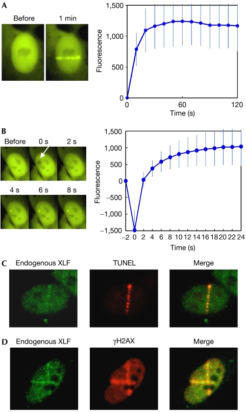
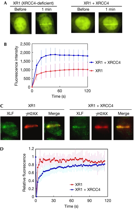
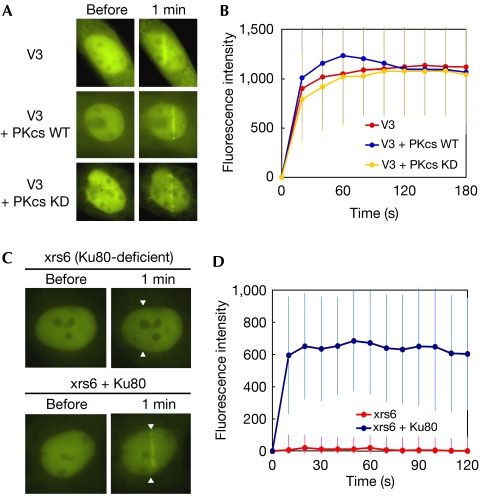
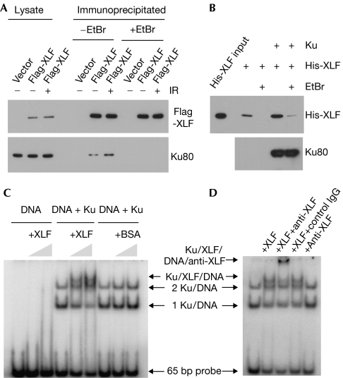
XRCC4-like factor (XLF)--also known as Cernunnos--has recently been shown to be involved in non-homologous end-joining (NHEJ), which is the main pathway for the repair of DNA double-strand breaks (DSBs) in mammalian cells. XLF is likely to enhance NHEJ by stimulating XRCC4-ligase IV-mediated joining of DSBs. Here, we report mechanistic details of XLF recruitment to DSBs. Live cell imaging combined with laser micro-irradiation showed that XLF is an early responder to DSBs and that Ku is essential for XLF recruitment to DSBs. Biochemical analysis showed that Ku-XLF interaction occurs on DNA and that Ku stimulates XLF binding to DNA. Unexpectedly, XRCC4 is dispensable for XLF recruitment to DSBs, although photobleaching analysis showed that XRCC4 stabilizes the binding of XLF to DSBs. Our observations showed the direct involvement of XLF in the dynamic assembly of the NHEJ machinery and provide mechanistic insights into DSB recognition.


Figures

Figure 1
Figure 1
Accumulation of XLF at laser-induced DNA double-strand breaks. (A) Accumulation of YFP-XLF at DSBs. Human 1BR3 cells expressing YFP-XLF were micro-irradiated across the nuclei. A typical example of the situation before and after (1 min) irradiation is shown (left). For kinetics analysis, the cell nucleus was exposed to a single burst of the laser, and images were obtained at 10 s intervals for 2 min. XLF accumulation at DSB sites was quantified and depicted in a graph (right). Mean values of the fluorescence intensities at each time point were calculated from ten independent measurements. (B) Rapid accumulation of YFP-XLF at DSBs. Images were taken at 2 s intervals. A typical example (left) and mean fluorescence intensities of ten independent measurements (right) are shown as in (A). The arrow indicates the site of micro-irradiation. (C) Colocalization of endogenous XLF protein and DSBs. Human 1BR3 cells were micro-irradiated and fixed. DSBs were labelled using TUNEL staining (red). Subsequently, the cells were stained with an XLF antibody (green). (D) Colocalization of XLF and γH2AX. 1BR3 cells were micro-irradiated, fixed and co-immunostained with XLF (green) and γH2AX (red) antibodies. DSB, double-strand break; TUNEL, TdT-mediated dUTP nick end labelling; XLF, XRCC4-like factor; YFP, yellow fluorescent protein; γH2AX, phosphorylated form of histone H2AX.
Figure 2
Figure 2
Dynamics of XLF in XRCC4-deficient and XRCC4-complemented cells. (A) Accumulation of YFP-XLF at DSBs. YFP-XLF was expressed in XRCC4-deficient XR1 (left) and XRCC4-complemented XR1 (right) cells. DSBs were generated by micro-irradiation and XLF accumulation was monitored before and 1 min after irradiation. (B) Kinetics of YFP-XLF accumulation at DSBs. Accumulation of YFP-XLF was measured as in Fig 1B. Mean values of the fluorescence intensities at each time point were calculated from ten independent measurements. (C) Accumulation of the endogenous XLF at DSBs in XR1 (left) and XRCC4-complemented (right) cells. Cells were micro-irradiated in their nucleus, fixed and stained with XLF and γH2AX antibodies. (D) FRAP analysis of YFP-XLF in XR1 and XRCC4-complemented XR1 cells. After maximum XLF accumulation, the whole DSB region was photobleached. Images were acquired before bleaching and at 1.5 s intervals after bleaching. Mean values of the fluorescence intensities at each time point were calculated from ten independent measurements. DSB, double-strand break; FRAP, fluorescence recovery after photobleaching; XLF, XRCC4-like factor; YFP, yellow fluorescent protein; γH2AX, phosphorylated form of histone H2AX.
Figure 3
Figure 3
XLF accumulation in DNA-PKCS-deficient and Ku80-deficient cells. (A) Accumulation of YFP-XLF at DSBs in DNA-PKCS-deficient (V3) cells (top), V3 cells complemented wild-type DNA-PKCS (middle) and V3 cells complemented with kinase-dead DNA-PKCS (bottom). Live cell imaging was carried out as described in Fig 1A. (B) Quantitative analysis of YFP-XLF accumulation at DSBs in DNA-PKCS-deficient and DNA-PKCS-complemented cells. Signal intensities were quantified and a mean value for each time point was calculated from ten independent measurements. (C) Accumulation of YFP-XLF at DSBs in Ku80-deficient xrs6 cells (top) and the Ku80-complemented xrs6 cells (bottom). Arrowheads indicate the sites of micro-irradiation. (D) Quantitative analysis of YFP-XLF accumulation at DSBs in xrs6 and Ku80-complemented xrs6. Images were obtained at 10 s intervals for 2 min. Signal intensities were quantified and a mean value with standard deviation at each time point was calculated from ten independent measurements. DNA-PKCS, DNA-dependent protein kinase catalytic subunit; DSB, double-strand break; KD, kinase dead; WT, wild type; XLF, XRCC4-like factor; YFP, yellow fluorescent protein.
Figure 4
Figure 4
Complex formation of XLF and Ku on DNA. (A) Ethidium bromide-sensitive co-precipitation of Ku with Flag-XLF. 1BR3 cells were transfected with Flag-XLF and treated with or without γ-rays at 10 Gy. Immunoprecipitation was carried out with a Flag antibody in the presence or absence of ethidium bromide, and the co-precipitation of Ku80 was analysed by western blotting. (B) Ku facilitates the XLF–DNA association. DNA-cellulose was reacted with or without purified Ku. The purified XLF was then incubated with DNA-cellulose with or without Ku. Ethidium bromide was added to the reactions as indicated. After washing, proteins bound on DNA-cellulose were analysed by western blotting. (C) EMSA of XLF and Ku. A 32P-labeled 65-bp DNA probe was reacted with purified Ku, XLF and BSA as indicated. Protein–DNA complexes were resolved by polyacrylamide gel electrophoresis and subsequently visualized by autoradiography. (D) Supershift assay using an XLF antibody. Ku and DNA probe were reacted with XLF, anti-XLF and control antibodies as indicated. BSA, bovine serum albumin; EtBr, ethidium bromide; DSB, double-strand break; EMSA, electrophoretic mobility shift assay; XLF, XRCC4-like factor; YFP, yellow fluorescent protein.

Similar articles

Cited by

References

    1. Ahnesorg P, Smith P, Jackson SP (2006) XLF interacts with the XRCC4–DNA ligase IV complex to promote DNA nonhomologous end-joining. Cell 124: 301–313 - PubMed
    1. Bryans M, Valenzano MC, Stamato TD (1999) Absence of DNA ligase IV protein in XR-1 cells: evidence for stabilization by XRCC4. Mutat Res 433: 53–58 - PubMed
    1. Buck D et al. (2006) Cernunnos, a novel nonhomologous end-joining factor, is mutated in human immunodeficiency with microcephaly. Cell 124: 287–299 - PubMed
    1. Callebaut I, Malivert L, Fischer A, Mornon JP, Revy P, de Villartay JP (2006) Cernunnos interacts with the XRCC4 x DNA–ligase IV complex and is homologous to the yeast nonhomologous end-joining factor Nej1. J Biol Chem 281: 13857–13860 - PubMed
    1. Calsou P, Delteil C, Frit P, Drouet J, Salles B (2003) Coordinated assembly of Ku and p460 subunits of the DNA-dependent protein kinase on DNA ends is necessary for XRCC4–ligase IV recruitment. J Mol Biol 326: 93–103 - PubMed

Publication types