. 2008 Jan;118(1):352-63.
doi: 10.1172/JCI32040.

Selective inhibition of proprotein convertases represses the metastatic potential of human colorectal tumor cells

Affiliations

Selective inhibition of proprotein convertases represses the metastatic potential of human colorectal tumor cells

Nathalie Scamuffa et al. J Clin Invest. 2008 Jan.

Abstract

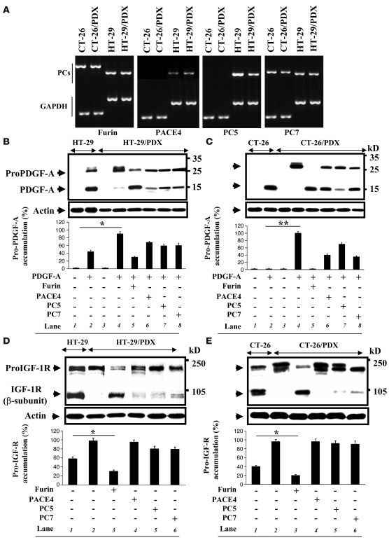
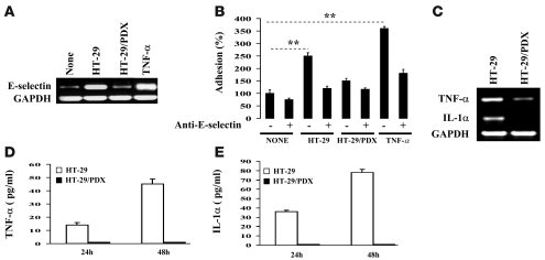
The proprotein convertases (PCs) are implicated in the activation of various precursor proteins that play an important role in tumor cell metastasis. Here, we report their involvement in the regulation of the metastatic potential of colorectal tumor cells. PC function in the human and murine colon carcinoma cell lines HT-29 and CT-26, respectively, was inhibited using siRNA targeting the PCs furin, PACE4, PC5, and PC7 or by overexpression of the general PC inhibitor alpha1-antitrypsin Portland (alpha1-PDX). We found that overexpression of alpha1-PDX and knockdown of furin expression inhibited processing of IGF-1 receptor and its subsequent activation by IGF-1 to induce IRS-1 and Akt phosphorylation, all important in colon carcinoma metastasis. These data suggest that the PC furin is a major IGF-1 receptor convertase. Expression of alpha1-PDX reduced the production of TNF-alpha and IL-1alpha by human colon carcinoma cells, and incubation of murine liver endothelial cells with conditioned media derived from these cells failed to induce tumor cell adhesion to activated murine endothelial cells, a critical step in metastatic invasion. Furthermore, colon carcinoma cells in which PC activity was inhibited by overexpression of alpha1-PDX when injected into the portal vein of mice showed a significantly reduced ability to form liver metastases. This suggests that inhibition of PCs is a potentially promising strategy for the prevention of colorectal liver metastasis.


Figures

Figure 1
Figure 1. Inhibition of PC activity in HT-29 and CT-26 colon cancer cells.
(A) RT-PCR analysis was performed on RNA extracted from the colon cancer cells HT-29 and CT-26 and the same cells that express the PC inhibitor α1-PDX (HT-29/PDX and CT-26/PDX) using furin, PACE4, PC5, PC7, and GAPDH-specific primers. All these PCs were present in HT-26 and HT-29/PDX cells, whereas CT-26 and CT-26/PDX cells lacked the expression of PC5 and PACE4. (BE) PDGF-A and IGF-1R processing was analyzed in the indicated cells by Western blotting using an anti-V5 (B and C) and anti–IGF-1R antibody (D and E), respectively. Note that α1-PDX inhibited the processing of these PC substrates, and only furin mediated complete rescue. Bars denote the corresponding percentages of pro-PDGF-A and pro-IGF-1R accumulation. Values are mean ± SEM (n = 3 per group). *P < 0.005; **P < 0.0002.
Figure 2
Figure 2. PC blockade inhibits IGF-1–induced IGF-1R, IRS-1, and Akt phosphorylation.
Serum-starved cells were stimulated for 10 minutes with 50 ng/ml IGF-1, and cell lysates were subjected to immunoprecipitation with either an anti–IGF-1R (A) or an anti–IRS-1 antibody (B). Immunoprecipitates were immunoblotted with anti-phosphotyrosine. (C) Cell lysates of IGF-1–activated cells were subjected to Western blotting with anti–phospho-Akt. The blots were stripped and reprobed with anti-actin or anti-Akt for data normalization. Results shown are representative of 3 experiments.
Figure 3
Figure 3. siRNA-mediated PC silencing on IGF-1R processing and IGF-1–induced Akt phosphorylation.
(A) RT-PCR analysis was performed on RNA extracted from HT-29 cells transfected with siRNA against furin, PC5, PACE4, or PC7. Note the downregulation of indicated PCs by their corresponding siRNA. (B) Western blotting analysis of IGF-1R processing in HT-29 cells transfected with the indicated siRNAs revealed that only siRNA against furin inhibited the processing of IGF-1R. (C) Serum-starved cells transfected with the indicated siRNAs were stimulated for 10 minutes with 50 ng/ml IGF-1 and cell lysates were subjected to Western blotting using anti–phospho-Akt. Note that only silencing of furin by siRNA significantly affected Akt phosphorylation. The blots were stripped and re-probed with anti-Akt for data normalization. Results shown are representative of 3 experiments.
Figure 4
Figure 4. Inhibition of PC activity alter E-selectin–dependent adhesion of tumor cells to endothelial cells and cytokine production.
(A) RT-PCR analysis was performed on RNA extracted from liver sinusoidal endothelial cells activated with TNF-α (10 ng/ml, positive control) or media derived from HT-29 or HT-29/PDX cells using E-selectin and GAPDH-specific primers. Note that media derived from HT-29/PDX cells failed to significantly induce E-selectin expression compared with media derived from HT-29 cells or TNF-α. (B) To test the effect of PC inhibition on tumor–endothelial cell adhesion, tumor cells were labeled with calcein and left to adhere to TNF-α (10 ng/ml) or tumor cell–derived, conditioned media–activated endothelial layer in the presence or absence of an anti–E-selectin neutralizing antibody. The attached tumor cells were quantified by measuring the fluorescence emission using a fluorometer. Only TNF-α and media derived from control cells significantly induced HT-29 adhesion. (C) RT-PCR analysis was performed on RNA extracted from HT-29 and HT-29/PDX cells using TNF-α, IL-1α, or GAPDH-specific primers. (D and E) Tumor cells were incubated at 37°C in serum-free media. After 24–48 hours, media were collected and analyzed for the presence of IL-1α and TNF-α using ELISA kit. Note the highly reduced expression and production of these cytokines in the presence of α1-PDX. Results shown are representative of 3 experiments. Data are mean ± SEM (n = 3 per group). **P < 0.001.
Figure 5
Figure 5. Induction of hepatic TNF-α mRNA by tumor cells.
(A and B) Mice were injected though the intrasplenic/portal route with 106 HT-29, HT-26/PDX (A), CT-26, or CT-26/PDX cells (B), and their livers were removed at the indicated times. Total RNA was extracted and analyzed by RT-PCR using specific primers for murine TNF-α or GAPDH. Results of laser densitometry are shown in the graph and are expressed as the ratio of TNF-α/GAPDH mRNA to that of control (liver of saline-injected mice at 1 hour), which was assigned a value of 1. In Panel A and B the time zero data is from a different RT-PCR experiment done at the same time. (C) Western blotting analysis of TNF-α expression in response to tumor cells 19 hours after injection. (D) Immunohistochemistry analysis of liver sections derived from the animals 19 hours following injection of tumor cells. The sections were stained with an anti-murine TNF-α antibody (green stain, denoted by white arrows). Yellow arrows indicate the lumen of liver vessels. Note that cells expressing α1-PDX failed to induce adequately hepatic TNF-α expression compared with control cells. No significant staining was observed in uninjected or saline-injected mice. Original magnification, ×40. Results shown are representative of 2 experiments. Values are mean ± SEM (n = 4 per group). *P < 0.05, **P < 0.001 versus respective α1-PDX–expressing group.
Figure 6
Figure 6. Hepatic E-selectin mRNA expression in response to tumor cells.
After the injections of 106 of CT-26, CT-26/PDX, HT-29, or HT-29/PDX cells, total RNA was extracted from mouse livers at different times and RT-PCR analysis was performed using E-selectin or GAPDH primers. Results of laser densitometry are shown in the bar graph and are expressed as the ratio of E-selectin/GAPDH mRNA to that of control (liver of saline-injected mice at 1 hour), which was assigned a value of 1. In Panel A and B the time zero data is from a different RT-PCR experiment done at the same time. (C) Western blotting analysis of E-selectin expression in response to tumor cells 19 hours after injection. (D) Immunohistochemistry analysis of liver sections derived from the animals 19 hours following injection of tumor cells. Sections were stained with an anti–E-selectin antibody (red stain, denoted by white arrows). Note that cells expressing α1-PDX failed to adequately induce hepatic E-selectin expression compared with control cells. Original magnification, ×40. Results shown are representative of 2 experiments. Values are mean ± SEM (n = 4 per group). *P < 0.05; **P < 0.001.
Figure 7
Figure 7. In vivo tumorigenicity assay.
HT-29, HT-29/PDX, CT-26, or CT-26/PDX cells (1 × 10⁶) were injected subcutaneously into 4-week-old female nude mice. The animals were monitored for tumor formation every 3 days. Results are representative of 4 experiments. Values are mean ± SEM (n = 6 per group). *P < 0.05; **P < 0.01.
Figure 8
Figure 8. PC inhibition prevents experimental liver metastasis and tumor vessel formation.
(A) Experimental liver metastases were generated by intrasplenic/portal injection of tumor cells, as described previously (–8). HT-29, HT-29/PDX, CT-26, or CT-26/PDX cells (1 × 10⁶) were injected into mice through intrasplenic/portal route, and liver metastases were enumerated 2 weeks after injection of CT-26 or CT-26/PDX cells and 4 weeks after injection of HT-29 or HT-29/PDX cells. (B) Developed liver tumors derived from HT-29– and HT-29/PDX–inoculated mice were analyzed for angiogenesis using an anti-mouse CD31 antibody. Original magnification, ×200.
Figure 9
Figure 9. PC activity in tumor cell–derived liver metastases.
(A) Indicated tumor cells were injected in mice through the intrasplenic/portal route. After 2 or 4 weeks, livers were removed and lysed in phosphate-buffered saline. Protein extracts were incubated with the intramolecularly quenched fluorogenic peptide Q-h-VEGF-C containing the PC processing site of VEGF-C, and the digestion of the QVEGF-C was evaluated as raw fluorescence units. (B) Aliquots of the enzymatic reaction reverse-phase HPLC chromatogram of the crude digest following incubation at 37°C of 20 μg QVEGF-C with liver protein extract. The elution of the peaks was monitored online by UV absorbance at 214 nm as well as by fluorescence detectors. (C) MALDI-ToF mass spectra of the crude digests following incubation of QVEGF-C with liver protein extract. The peak at m/z 1,703 is attributed to QVEGF-C. The peaks at m/z 1,127 and 594 were attributed to the highly fluorescent N-terminal [NT; Abz-Q-VHSIIRR-OH] and the nonfluorescent C-terminal [CT; SLP(NO2)-A-CONH2] fragments, respectively. (D) Immunoblotting analysis of IGF-1R processing in protein extracts derived from indicated tumor cell–derived metastases revealed the inhibition of IGF-1R processing in CT-26/PDX–derived metastases. Results are representative of 3 experiments. Values are mean ± SEM (n = 3 per group). **P < 0.001.

References

    1. Choti M.A., Bulkley G.B. Management of hepatic metastases. Liver Transpl. Surg. 1999;5:65–80. - PubMed
    1. Hiratsuka M., et al. Management of patients with hepatic metastases from gastric carcinoma. Nippon Geka Gakkai Zasshi. 2003;104:711–716. - PubMed
    1. Jaeck D. The significance of hepatic pedicle lymph nodes metastases in surgical management of colorectal liver metastases and of other liver malignancies. Ann. Surg. Oncol. 2003;10:1007–1011. - PubMed
    1. Vallet C., Martinet O., Mosimann F. Surgical treatment of hepatic metastases. Rev. Med. Suisse Romande. 2001;121:119–124. - PubMed
    1. Kitakata H., et al. Essential roles of tumor necrosis factor receptor p55 in liver metastasis of intrasplenic administration of colon 26 cells. Cancer Res. 2002;62:6682–6687. - PubMed

Publication types

MeSH terms