. 2009 Feb;30(2):432-51.
doi: 10.1002/hbm.20514.

Neurofunctional modulation of brain regions by distinct forms of motor cognition and movement features

Affiliations

Neurofunctional modulation of brain regions by distinct forms of motor cognition and movement features

Martina Piefke et al. Hum Brain Mapp. 2009 Feb.

Abstract

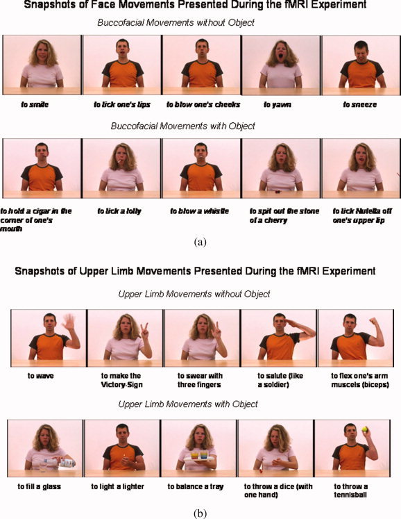
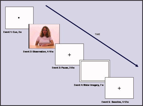
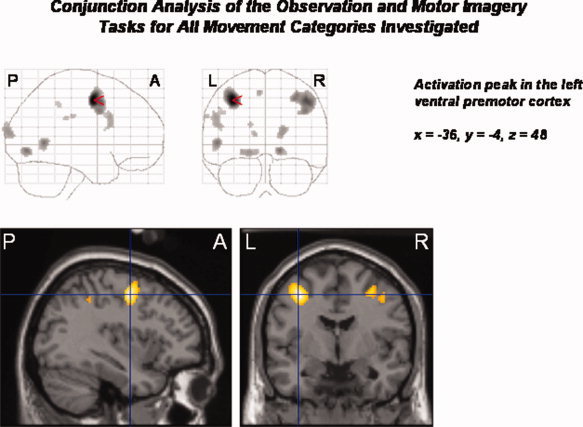
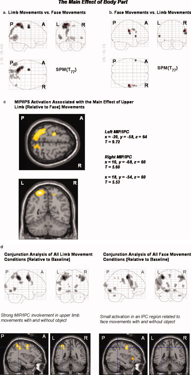
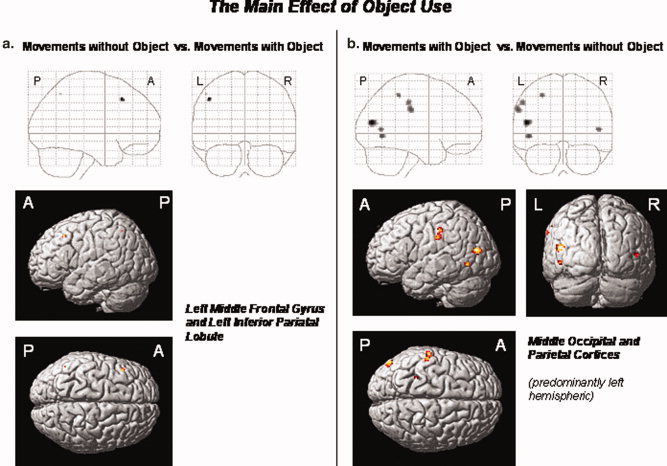
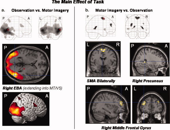
Extrastriate, parietal, and frontal brain regions are differentially involved in distinct kinds of body movements and motor cognition. Using functional magnetic resonance imaging, we investigated the neural mechanisms underlying the observation and mental imagery of meaningful face and limb movements with or without objects. The supplementary motor area was differentially recruited by the mental imagery of movements while there were differential responses of the extrastriate body area (EBA) during the observation conditions. Contrary to most previous reports, the EBA responded to face movements, albeit to a lesser degree than to limb movements. The medial wall of the intraparietal sulcus and adjacent intraparietal cortex was selectively recruited by the processing of meaningful upper limb movements, irrespective of whether these were object-related or not. Besides reach and grasp movements, the intraparietal sulcus may thus be involved in limb gesture processing, that is, in an important aspect of human social communication. We conclude that subregions of a frontal-parietal network differentially interact during the cognitive processing of body movements according to the specific motor-related task at hand and the particular movement features involved.


Figures

Figure 1
Figure 1
Examples of the meaningful body movements shown in video clips during the fMRI experiment. Half of the movements of each stimulus category—buccofacial movements either with or without object (a) and upper limb movements either with or without object (b)—were performed by a male, and the other half by a female.
Figure 2
Figure 2
Order and duration of events included in each experimental trial. The cue event at the beginning was always followed by an observation (video clip), a pause (fixation cross), a motor imagery (motor imagery of the movement seen in the preceding video clip), and a “baseline” event (fixation cross). Event duration was jittered to prevent correlation of event regressors. A reaction time task was included in the experiment to control for the subjects' alertness during the experiment (button press response upon detection of a checkerboard presented for 500 ms in 50% of the baseline events).
Figure 3
Figure 3
Conjunction analysis of all experimental conditions of interest (observation and motor imagery of buccofacial movements without object, buccofacial movements with object, upper limb movements without object, upper limb movements with object; relative to the baseline condition). There is increased neural activity in a bilateral network of frontal, parietal, occipital, and extrastriate brain areas, including the precentral and inferior frontal gyri, the SMA, the inferior parietal lobule, lingual, supramarginal, and superior occipital gyri, the cuneus, and the EBA. Activations predominated in the left hemisphere. The activation peak was located in the left superior PMv. Statistical threshold: P < 0.05, FWE corrected across the whole brain volume (T = 5.11); k = 0 voxel. P, posterior; A, anterior; L, left; R, right.
Figure 4
Figure 4
(a–d) Main effect of the factor “body part” and separate conjunction analyses for face and upper limb movements (each with and without object): The processing of limb (relative to face) movements bilaterally activated area MIP and the adjacent IPC, the EBA, calcarine sulci, and lingual gyri (extending into the fusiform gyrus in the left hemisphere), the left postcentral gyrus/inferior parietal lobule, superior/middle frontal, and superior occipital gyri, as well as the right precuneus. The reverse contrast (the processing of face relative to limb movements) revealed differential neural responses in the postcentral and inferior frontal gyri bilaterally, as well as the right supramarginal and precentral gyri (a–c). A conjunction analysis of the limb conditions (relative to baseline) showed that both limb movements with and without object reference activated the left MIP and adjacent IPC. A respective conjunction analysis of the face conditions revealed only small bilateral IPC activations in distinct anatomical regions (d). Statistical threshold: P < 0.05, FWE corrected across the whole brain volume (T = 5.11); k = 0 voxel. P, posterior; A, anterior; L, left; R, right.
Figure 5
Figure 5
(a,b) The main effect of the factor “object use”: The processing of movements without object (relative to object‐related movements) differentially activated the left middle frontal gyrus and inferior parietal lobule (a). The reverse contrast (object‐related versus nonobject‐related movements) revealed differential neural responses in the EBA and adjacent area MT/V5 bilaterally, the left middle occipital and supramarginal gyri, and the left inferior parietal lobule (b). Statistical threshold: P < 0.05, FWE corrected across the whole brain volume (T = 5.11); k = 0 voxel. P, posterior; A, anterior; L, left; R, right.
Figure 6
Figure 6
(a,b) The main effect of the factor “task”: The observation (relative to the mental imagery) of all movement types differentially activated the right EBA, the inferior occipital gyrus bilaterally, the right inferior frontal gyrus, intraparietal sulcus and adjacent IPC, thalamus, and cerebellum (a). The reverse contrast (motor imagery relative to the observation of all movement types) revealed differential activations of the right precuneus, SMA and middle frontal gyri bilaterally, the left rolandic operculum and posterior cingulate cortex (extending into the hippocampal region), as well as the right insula (extending into the temporal pole) (b). Statistical threshold: P < 0.05, FWE corrected across the whole brain volume (T = 5.11); k = 0 voxel. P, posterior; A, anterior; L, left; R, right.

Similar articles

Cited by

References

    1. Assmus A,Marshall JC,Ritzl A,Noth J,Zilles K,Fink GR ( 2003): Left inferior parietal cortex integrates time and space during collision judgements. Neuroimage 20 ( Suppl 1): S82–S88. - PubMed
    1. Assmus A,Giessing C,Weiss PH,Fink GR ( 2007): Functional interactions during the retrieval of conceptual action knowledge: An fMRI study. J Cogn Neurosci 19: 1004–1012. - PubMed
    1. Astafiew SV,Stanley CM,Shulman GL,Corbetta M ( 2004): Extrastriate body area in human occipital cortex responds to the performance of motor actions. Nat Neurosci 7: 422–423. - PubMed
    1. Aziz‐Zadeh L,Iacoboni M,Zaidel E ( 2006a): Hemispheric sensitivity to body stimuli in simplke reaction time. Exp Brain Res 170: 116–121. - PubMed
    1. Aziz‐Zadeh L,Koski L,Zaidel E,Mazziotta J,Iacoboni M ( 2006b): Lateralization of the human mirror neuron system. J Neurosci 26: 2964–2970. - PMC - PubMed

Publication types

MeSH terms