. 2008 Jun;215(3):715-24.
doi: 10.1002/jcp.21354.

Epac/Rap and PKA are novel mechanisms of ANP-induced Rac-mediated pulmonary endothelial barrier protection

Affiliations

Epac/Rap and PKA are novel mechanisms of ANP-induced Rac-mediated pulmonary endothelial barrier protection

Anna A Birukova et al. J Cell Physiol. 2008 Jun.

Abstract

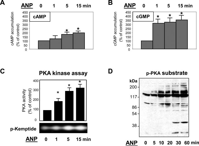
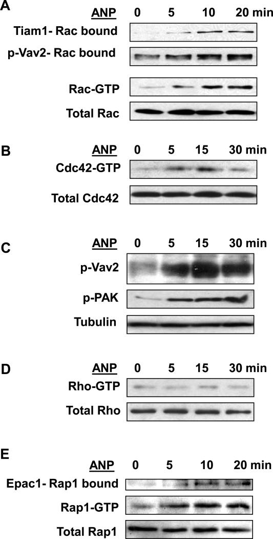
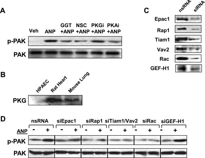
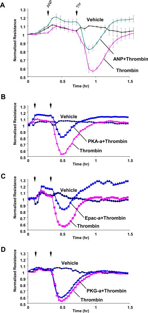
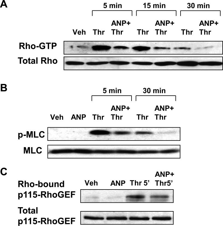
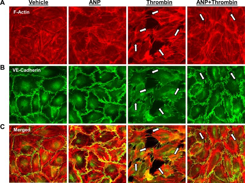
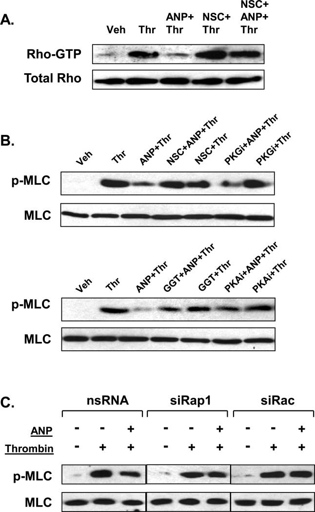
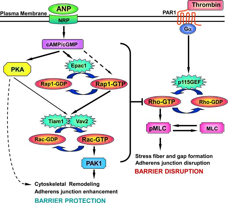
Acute lung injury, sepsis, lung inflammation, and ventilator-induced lung injury are life-threatening conditions associated with lung vascular barrier dysfunction, which may lead to pulmonary edema. Increased levels of atrial natriuretic peptide (ANP) in lung circulation reported in these pathologies suggest its potential role in the modulation of lung injury. Besides well recognized physiological effects on vascular tone, plasma volume, and renal function, ANP may exhibit protective effects in models of lung vascular endothelial cell (EC) barrier dysfunction. However, the molecular mechanisms of ANP protective effects are not well understood. The recently described cAMP-dependent guanine nucleotide exchange factor (GEF) Epac activates small GTPase Rap1, which results in activation of small GTPase Rac-specific GEFs Tiam1 and Vav2 and Rac-mediated EC barrier protective responses. Our results show that ANP stimulated protein kinase A and the Epac/Rap1/Tiam/Vav/Rac cascade dramatically attenuated thrombin-induced pulmonary EC permeability and the disruption of EC monolayer integrity. Using pharmacological and molecular activation and inhibition of cAMP-and cGMP-dependent protein kinases (PKA and PKG), Epac, Rap1, Tiam1, Vav2, and Rac we linked ANP-mediated protective effects to the activation of Epac/Rap and PKA signaling cascades, which dramatically inhibited the Rho pathway of thrombin-induced EC hyper-permeability. These results suggest a novel mechanism of ANP protective effects against agonist-induced pulmonary EC barrier dysfunction via inhibition of Rho signaling by Epac/Rap1-Rac and PKA signaling cascades.


Figures

Figure 1
Figure 1. Effect of ANP on cAMP, cGMP, and PKA activation
A and B: EC were stimulated with 100nM of ANP for indicated periods of time, and intracellular cAMP (Panel A) and cGMP (Panel B) levels were determined using a non-radioactive immunoassay, as described in Materials and Methods. Results are mean ± SD of three independent experiments. *P<0.001. C: Cell lysates were analyzed for PKA activity by non-radioactive in vitro PKA assay. The inset represents fluorescent phosphorylated form of PKA substrate kemptide separated from non-phosphorylated form by 0.8% agarose gel electrophoresis. The fluorescence intensity was detected and quantified by a EagleEye Image System. Results are mean ± SD of three independent experiments. *P<0.001. D: PKA-mediated phosphorylation of endogenous substrates was monitored by immunoblotting with anti-phospho-PKA substrate antibody.
Figure 2
Figure 2. Effect of ANP on activation of small GTPases Rac and Rac-dependent signaling, Cdc42, Rho, and Rap1
EC were stimulated with 100nM of ANP for indicated periods of time. A: Results of Rac activation assay. Upper panels depict Tiam1 and p-Vav2 bound to activate Rac (Rac-GTP). Middle panel depicts the levels of activated Rac, and the lower panel shows total Rac content in EC lysates. B: Results of Cdc42 activation assay. Upper panel depicts the levels of activated Cdc42, and the lower panel shows total Cdc42 content in EC lysates. C: Phosphorylation of Vav2 and PAK1 in control and ANP-stimulated EC was determined in the total lysates using specific antibodies. Equal loading was confirmed by probing of membranes for β-tubulin. D: Results of Rho activation assay. Upper panel depicts the levels of activated Rho, and the lower panel shows total Rho content in EC lysates. E: Rap1 activation pull-down assay. Upper panels depict Epac1 bound to activate Rap1 (Rap-GTP). Lower panel shows total Rap1 content in EC lysates. Results are representative of three independent experiments.
Figure 3
Figure 3. Involvement of Epac/Rap1-, Tiam1-Vav2- and PKA-dependent mechanisms in the regulation ANP-mediated Rac activation
A: EC were pretreated with NSC-23766 (200μM), PKI (10μM), GGTI-298 (10μM), or Rp-8-Br-cGMPS (10μM) for 30 min followed by stimulation with ANP (100 nM, 15 min). Phosphorylation of PAK1 was determined in the total lysates using phospho-PAK1 specific antibody. Results are representative of three independent experiments. B: Expression of PKG has been analyzed by western blot in samples obtained from HPAEC, rat heart, and mouse lung. C: Pulmonary EC were transfected with siRNA specific to Epac1, Rap1, Tiam1, Vav2, Rac, or GEF-H1. Depletion of target proteins induced by specific siRNA duplexes was confirmed by immunoblotting with appropriate antibody, as compared to treatment with non-specific RNA. Results are representative of three to five independent experiments. D: Pulmonary EC were transfected with specific siRNAs followed by ANP stimulation and detection of PAK1 phosphorylation using specific antibody. Control cells were treated with non-specific RNA. Results are representative of three independent experiments.
Figure 4
Figure 4. Effect of ANP, PKA, Epac, and PKG activators on thrombin-induced permeability
Human pulmonary EC were treated with ANP (100 nM, marked by first arrow). At the time point indicated by second arrow, cells were stimulated with thrombin (0.5 U/ml), and TER was monitored over the time (upper panel) (A). In complementary experiments N6-Benzoyl-cAMP (100μM) (B), 8-CPT-2’-O-Me-cAMP (100μM) (C), or SP-8-Br-PET-cGMPS (100μM) (D) were used instead of ANP.
Figure 5
Figure 5. Effect of ANP on thrombin-induced modulation of Rho- and Rac-dependent pathways
Pulmonary EC were pre-incubated with ANP (100nM, 15 min), followed by treatment with thrombin (0.5 U/ml) for 5 min, 15 min or 30 min.A: Rho activation pull-down assay. Upper panel depicts the levels of activated Rho, and the lower panel shows total Rho content in EC lysates. B: Phosphorylation of MLC in EC pretreated with ANP followed by thrombin challenge was detected by western blot with diphospho-MLC specific antibodies. The lower panel represents the membrane re-probed with pan-MLC antibody. C: The upper panel depicts p115Rho-GEF associated with activated Rho. The lower panel represents p115Rho-GEF content in the total lysates detected by western blot. Results in each group are representative of three independent experiments.
Figure 6
Figure 6. Effect of ANP on thrombin-induced EC remodeling of actin cytoskeleton, adherens junctions and monolayer disruption
A and B: EC grown on glass coverslips were preincubated with ANP (100nM 15 min), followed by thrombin treatment (0.5 U/ml) for 15 min and double immunofluorescence staining with Texas Red phalloidin to detect actin filaments (A) and with VE-cadherin antibodies to visualize adherens junctions (B). C: Merged images depict ANP-induced accumulation of peripheral F-actin at adherens junctions’ areas and ANP-mediated preservation of EC monolayer integrity against thrombin-induced disruption. Paracellular gaps and disrupted intercellular contacts are marked by arrows. Results are representative of three independent experiments.
Figure 7
Figure 7. Effects of Rac, Rap1, and PKA inhibition on modulation of thrombin-induced Rho signaling by ANP
A: EC preincubated with vehicle or NSC-23766 (200 μM, 30min), were treated with ANP (100nM, 15 min) and stimulated with thrombin (0.5 U/ml, 15 min). Control cells were treated with thrombin alone. Rho activation pull-down assays were performed as described in Methods. B: HPAEC were pretreated with NSC-23766 (200μM), Rp-8-Br-cGMPS (10μM) (upper panels), GGTI-298 (10μM), or PKI (10μM) (lower panels) for 30 min followed by ANP stimulation (100nM, 15 min) and thrombin challenge (0.5 U/ml, 15 min). Phosphorylation of MLC was determined in the total lysates using phospho-specific antibodies. Results are representative of three independent experiments. C: Pulmonary EC transfected with Rap1- or Rac-specific siRNA or non-specific RNA duplexes were stimulated with vehicle or ANP (100 nM, 15 min), followed by thrombin addition (0.5 U/ml, 15 min). Phosphorylated MLC was determined by immunoblotting using MLC phospho-specific antibodies. The lower panel represents the membrane re-probed with pan-MLC antibody.
Figure 8
Figure 8. Upstream mechanisms of ANP-induced Rac activation and modulation of Rho pathway of EC barrier dysfunction
Stimulation of EC with ANP elevates intracellular cAMP/cGMP levels and stimulates cAMP-dependent protein kinase (PKA) and cAMP-activated guanine nucleotide exchange factor Epac1, which activates its effector small GTPase Rap1. Epac1 may be also activated by high local cGMP concentrations (Christensen et al., 2003). In addition, Rap1 may be activated by putative cGMP-specific GEF (von Lintig et al., 2000). Activated PKA and Rap1 promote Rac activation via stimulation of Rac specific GEFs Tiam1 and Vav2. Activated Rac attenuates the Rho pathway of endothelial barrier dysfunction via reduction of Rho activity, which leads to decreased myosin light chain phosphorylation, EC contraction, and less severe endothelial barrier dysfunction. In addition, PKA may directly affect EC cytoskeletal organization and monolayer barrier properties via modulation of myosin light chain kinase activity or VASP-dependent relaxation of actin cytoskeleton.

References

    1. Aghazadeh B, Lowry WE, Huang XY, Rosen MK. Structural basis for relief of autoinhibition of the Dbl homology domain of proto-oncogene Vav by tyrosine phosphorylation. Cell. 2000;102(5):625–633. - PubMed
    1. Ahluwalia A, MacAllister RJ, Hobbs AJ. Vascular actions of natriuretic peptides. Cyclic GMP-dependent and -independent mechanisms. Basic Res Cardiol. 2004;99(2):83–89. - PubMed
    1. Arthur WT, Quilliam LA, Cooper JA. Rap1 promotes cell spreading by localizing Rac guanine nucleotide exchange factors. J Cell Biol. 2004;167(1):111–122. - PMC - PubMed
    1. Baxter GF. The natriuretic peptides. Basic Res Cardiol. 2004;99(2):71–75. - PubMed
    1. Birukov KG, Bochkov VN, Birukova AA, Kawkitinarong K, Rios A, Leitner A, Verin AD, Bokoch GM, Leitinger N, Garcia JG. Epoxycyclopentenone-containing oxidized phospholipids restore endothelial barrier function via Cdc42 and Rac. Circ Res. 2004;95(9):892–901. - PubMed

MeSH terms

LinkOut - more resources