. 2008 Apr 15;94(8):3003-13.
doi: 10.1529/biophysj.107.123448. Epub 2007 Dec 7.

Pore mutations of the Escherichia coli MscS channel affect desensitization but not ionic preference

Affiliations

Pore mutations of the Escherichia coli MscS channel affect desensitization but not ionic preference

Michelle D Edwards et al. Biophys J. .

Abstract

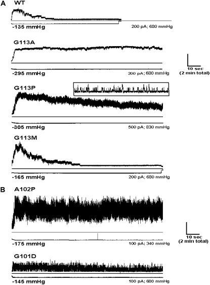
Mechanosensitive channels rescue bacterial cells from a fate of lysis when they transfer from a high- to low-osmolarity environment. Of three Escherichia coli mechanosensitive proteins studied to date, only MscS-Ec demonstrates a small anionic preference and a desensitized, nonconducting state under sustained pressure. Little is known about the mechanisms generating these distinctive properties. Eliminating the sole positive charge in the MscS-Ec pore region (Arg(88)) did not alter anionic preference. Adding positive charges at either end of the pore did not augment anionic preference, and placing negative charges within the pore did not diminish it. Thus, pore charges do not control this characteristic. However, from this analysis we identified mutations in the hinge region of the MscS-Ec pore helix (at Gly(113)) that profoundly affected ability of the channel to desensitize. Substitution with nonpolar (Ala, Pro) or polar (Asp, Arg, Ser) residues inhibited transition to the desensitized state. Interestingly, Gly(113) replaced with Met did not impede desensitization. Thus, although Gly is not specifically required at position 113, MscS desensitization is strongly influenced by the residue situated here. Mutations at residues further into the pore also regulated desensitization. Transition to this unique mechanosensitive channel state is discussed in terms of existing data.


Figures

FIGURE 1
FIGURE 1
Location of pore mutations in the MscS-Ec crystal structure. The heptameric structure of MscS-Ec (30) is depicted in full length as a side view (left panel) and as viewed from the top (bottom right), and a zoomed-in view of the pore region for two subunits is also shown (residues 80–127, chains B and E; top right). Each subunit backbone is drawn with residues mutated in this study highlighted by space filling. (Images created in Chime (32).)
FIGURE 2
FIGURE 2
Membrane expression of mutant MscS proteins. Mutant channels were overexpressed in MJF465 cells (30 min 0.3 mM IPTG), and membrane protein samples were extracted, separated by SDS-PAGE, transferred to nitrocellulose membrane, and probed with anti-YggB antibody. (Left-hand blot) 1, markers; 2, MJF465 cells containing no plasmid; 3, pMscSH6 (His-tagged wild-type MscS); 4, R88S pMscSH6; 5, T93R pMscSH6; 6, G101D pMscSH6. (Right-hand blot) 1, markers; 2, MJF465 cells containing no plasmid; 3, pMscSH6 (His-tagged wild-type MscS); 4, G113A pMscS; 5, G113D pMscSH6; 6, G113M pMscS; 7, G113P pMscS; 8, G113R pMscSH6; 9, G113S pMscS. Note that some of the G113 mutants were constructed in a non-His plasmid, and accordingly, these mutant proteins run slightly faster on the gel (lanes 4, 6, 7, and 9).
FIGURE 3
FIGURE 3
Current-voltage properties of mutant MscS channels. Protoplasts containing either wild-type or mutant MscS channels were prepared. Excised patches were bathed in symmetrical solutions (200 mM KCl), and channel activity was recorded from holding potentials of −50 mV to +50 mV (10-mV steps). The bath solution was then replaced with buffer containing 600 mM KCl, and channel activity was recorded again at the same potentials. Single-channel current is plotted against membrane voltage for both sets of recording conditions (mean ± SE). (A) Plots for wild-type (WT) MscS and single-point mutants with a positively charged Arg either removed or inserted. (B) Plots for single-point mutants where a negatively charged Asp has been added to the pore region. (C) The mean current (+ SE) measured at +50 mV in symmetrical solutions is compared for each channel. Only G113R channels exhibited significantly lower levels of current compared with wild-type channels (p < 0.05, Student's t-test). (D) The deletion mutant Δ266–286, where the last 21 amino acids are absent from each subunit, shows no significant change in reversal potential in asymmetric solutions; however, this mutant channel exhibits a much larger conductance than wild-type channels at positive membrane potentials in both symmetrical and asymmetric solutions.
FIGURE 4
FIGURE 4
Mutation G113R induces visits to substates at lower membrane potentials. Zoomed-in sections of traces illustrating single channel openings for (A) wild-type (WT) and (B) G113R MscS channels at +20 mV membrane potential. Wild-type channels tend to open or close at this voltage and rarely visit subconducting states; however, G113R channels flicker in and out of substates. Usually this occurs rapidly (see examples i and ii), but sometimes the protein will remain in the subconducting conformation for tens of milliseconds (see example ii, arrow).
FIGURE 5
FIGURE 5
Bulky, polar substitutions at residue 113 inhibit desensitization of MscS-Ec. Proteins containing a substitution at position 113 to Asp, Arg, or Ser were expressed in MJF429 cells, and protoplasts were prepared. Pressure was applied to excised membrane patches and clamped once all MscS channels had opened. Channel activity was followed under the maintained stimulus for up to 4 min, and 2 min of representative example recordings are depicted. For each example, traces show total current (channel activity) in upper panel and pressure level in lower panel (the actual pressure applied for each example is indicated under the pressure trace). Wild-type channels desensitize fully within this time, but G113D, G113R, and G113S MscS channels continue to close and reopen for as long as pressure is held.
FIGURE 6
FIGURE 6
Mutations at a number of TM3 sites block MscS-Ec desensitization. Experiments were carried out as described for Fig. 5, and representative current and pressure traces are similarly depicted. (A) Gly113 is not essential for desensitization. Although substitutions G113A and G113P removed the ability of the protein to enter the desensitized state, mutation G113M had no such effect. Placement of Pro at 113 shortened channel open dwell times, producing a flickery channel activity. Inset illustrates these single channel openings at an increased time scale. (B) Mutations at Ala102 and Gly101 also eliminated the capacity for desensitization. As detailed in the main text, A102P and G101D channels gated with shorter open dwell times, thus producing the flickery activity observed in their current traces. Also, A102P channels exhibited smaller conductance—about half of wild-type MscS—and thus the figure depicts a patch where ∼10–20 channels are open at any one time.

References

    1. Martinac, B., M. Buehner, A. H. Delcour, J. Adler, and C. Kung. 1987. Pressure-sensitive ion channel in Escherichia coli. Proc. Natl. Acad. Sci. USA. 84:2297–2301. - PMC - PubMed
    1. Berrier, C., A. Coulombe, C. Houssin, and A. Ghazi. 1989. A patch-clamp study of ion channels of inner and outer membranes and of contact zones of E. coli, fused into giant liposomes. FEBS Lett. 259:27–32. - PubMed
    1. Sukharev, S. I., B. Martinac, V. Y. Arshavsky, and C. Kung. 1993. Two types of mechanosensitive channels in the Escherichia coli cell envelope: solubilization and functional reconstitution. Biophys. J. 65:177–183. - PMC - PubMed
    1. Blount, P., S. I. Sukharev, P. C. Moe, S. K. Nagle, and C. Kung. 1996. Towards an understanding of the structural and functional properties of MscL, a mechanosensitive channel in bacteria. Biol. Cell. 87:1–8. - PubMed
    1. Moe, P. C., P. Blount, and C. Kung. 1998. Functional and structural conservation in the mechanosensitive channel MscL implicates elements crucial for mechanosensation. Mol. Microbiol. 28:583–592. - PubMed

Publication types

LinkOut - more resources