. 2008 Feb;190(4):1459-72.
doi: 10.1128/JB.01688-07. Epub 2007 Dec 7.

Moraxella catarrhalis synthesizes an autotransporter that is an acid phosphatase

Affiliations

Moraxella catarrhalis synthesizes an autotransporter that is an acid phosphatase

Todd C Hoopman et al. J Bacteriol. 2008 Feb.

Abstract

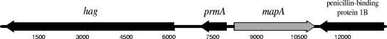
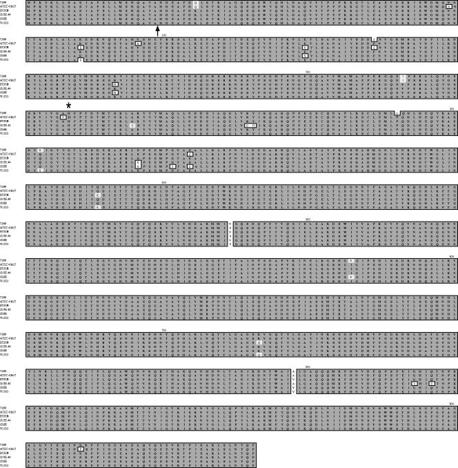
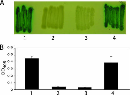
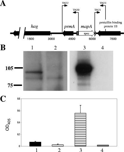
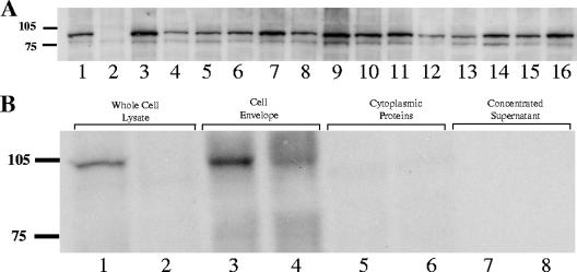
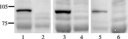
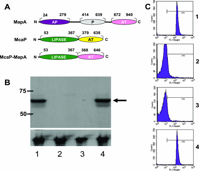
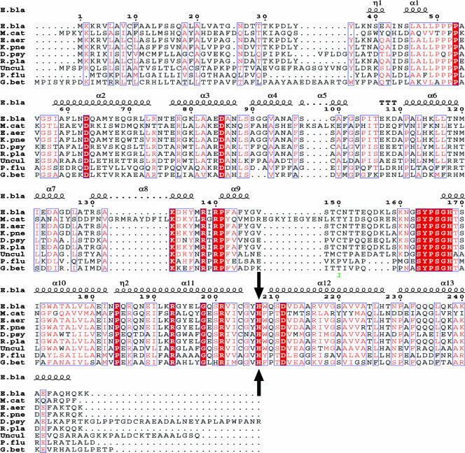
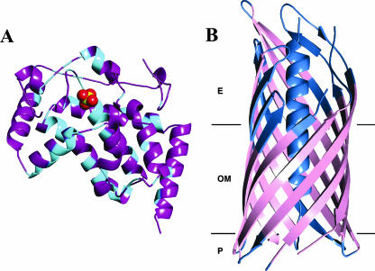
Moraxella catarrhalis O35E was shown to synthesize a 105-kDa protein that has similarity to both acid phosphatases and autotransporters. The N-terminal portion of the M. catarrhalis acid phosphatase A (MapA) was most similar (the BLAST probability score was 10(-10)) to bacterial class A nonspecific acid phosphatases. The central region of the MapA protein had similarity to passenger domains of other autotransporter proteins, whereas the C-terminal portion of MapA resembled the translocation domain of conventional autotransporters. Cloning and expression of the M. catarrhalis mapA gene in Escherichia coli confirmed the presence of acid phosphatase activity in the MapA protein. The MapA protein was shown to be localized to the outer membrane of M. catarrhalis and was not detected either in the soluble cytoplasmic fraction from disrupted M. catarrhalis cells or in the spent culture supernatant fluid from M. catarrhalis. Use of the predicted MapA translocation domain in a fusion construct with the passenger domain from another predicted M. catarrhalis autotransporter confirmed the translocation ability of this MapA domain. Inactivation of the mapA gene in M. catarrhalis strain O35E reduced the acid phosphatase activity expressed by this organism, and this mutation could be complemented in trans with the wild-type mapA gene. Nucleotide sequence analysis of the mapA gene from six M. catarrhalis strains showed that this protein was highly conserved among strains of this pathogen. Site-directed mutagenesis of a critical histidine residue (H233A) in the predicted active site of the acid phosphatase domain in MapA eliminated acid phosphatase activity in the recombinant MapA protein. This is the first description of an autotransporter protein that expresses acid phosphatase activity.


Figures

FIG. 1.
FIG. 1.
Schematic representation of the M. catarrhalis ATCC 43617 locus containing the mapA gene and adjacent genes, including hag.
FIG. 2.
FIG. 2.
Alignment of the predicted amino acid sequence of the MapA proteins from M. catarrhalis strains 7169, ATCC 43617, ETSU-9, O12E.44, O35E, and V1120. The arrow indicates the predicted signal peptidase I cleavage site between aa 23 and 24. The asterisk indicates the position of the histidine residue (aa 233) located in the active site of the acid phosphatase domain (described in Results).
FIG. 3.
FIG. 3.
Expression of acid phosphatase activity by recombinant E. coli cells. (A) Appearance of recombinant E. coli strains grown overnight on phenolphthalein-methyl green agar. Lanes: 1, E. coli DH5α(pKW04); 2, E. coli DH5α(pTH10); 3, E. coli DH5α(pTH24) with the H233A mutation; 4, E. coli DH5α(pTH26) with the H110A mutation. (B) Acid phosphatase assay using PnPP as the substrate with whole cells (2.5 × 10⁶ CFU) of E. coli DH5α(pKW04) (bar 1), E. coli DH5α(pTH10) (bar 2), E. coli DH5α(pTH24) (bar 3), and E. coli DH5α(pTH26) (bar 4). The data represent the means from three independent experiments, with error bars showing the standard deviations of the means. OD405, optical density at 405 nm.
FIG. 4.
FIG. 4.
Construction and characterization of an M. catarrhalis mapA mutant. (A) Schematic showing the mapA deletion in the chromosome of the M. catarrhalis O35EΔmapA mutant, together with the relevant primers used for PCR (with wild-type O35E chromosomal DNA) and overlapping extension PCR. (B) Western blot analysis of MapA expression by whole-cell lysates of O35E (lane 1), O35EΔmapA (lane 2), O35EΔmapA-9(pTH13) (lane 3), and O35EΔmapA-9(pWW115) (lane 4). All four samples were run on the same gel; exposure times different from those used for lanes 1 and 2 were used for lanes 3 and 4. Molecular mass position markers (in kilodaltons) are presented on the left side of these panels. (C) Acid phosphatase assay using PnPP as the substrate with whole cells (5 × 10⁸ CFU) of wild-type M. catarrhalis O35E (bar 1) and the O35EΔmapA mutant (bar 2) and with whole cells (5 × 10⁶ CFU) of M. catarrhalis O35EΔmapA-9(pTH13) (bar 3) and the O35EΔmapA-9(pWW115) (bar 4). The data in panel C represent the means from three independent experiments, with error bars showing the standard deviations of the means. OD405, optical density at 405 nm.
FIG. 5.
FIG. 5.
Detection of the MapA protein in other M. catarrhalis strains and in different cell fractions. (A) Western blot analysis using the MapA-directed MAb 1H12 to probe whole-cell lysates of the following M. catarrhalis strains: O35E (lane 1), the O35EΔmapA mutant (lane 2), FIN2265 (lane 3), FIN2406 (lane 4), FIN2344 (lane 5), V1145 (lane 6), V1156 (lane 7), V1120 (lane 8), FR3221 (lane 9), FR2213 (lane 10), FR2336 (lane 11), B59911 (lane 12), B59504 (lane 13), ETSU-17 (lane 14), ETSU-5 (lane 15), and ATCC 43617 (lane 16). (B) Western blot analysis using the MapA-directed MAb 1H12 to probe whole-cell lysates (lanes 1 and 2), cell envelopes (lanes 3 and 4), cytoplasmic proteins (lanes 5 and 6), and concentrated culture supernatant fluid (lanes 7 and 8) prepared from M. catarrhalis O35E (lanes 1, 3, 5, and 7) and the O35EΔmapA mutant (lanes 2, 4, 6, and 8). Protein loads in each pair of lanes 3 to 8 were standardized by means of the Bradford protein assay. Molecular mass position markers (in kilodaltons) are present on the left side of the figure.
FIG. 6.
FIG. 6.
Detection of MapA in outer membrane vesicles from three M. catarrhalis strains. Proteins present in outer membrane vesicles prepared from O35E (lane 1), the O35EΔmapA mutant (lane 2), ETSU-9 (lane 3), the ETSU-9ΔmapA mutant (lane 4), 7169 (lane 5), and the 7169ΔmapA (lane 6) mutant were resolved by SDS-PAGE and probed by Western blot analysis with MAb 1H12. The Bradford method was used to standardize protein amounts. Molecular mass position markers (in kilodaltons) are present on the left side of the figure.
FIG. 7.
FIG. 7.
Construction and characterization of an McaP-MapA fusion protein. (A) Schematic of the predicted domains used to produce the McaP-MapA fusion protein expressed by pTH34. AP, acid phosphatase domain in MapA; P, pertactin domain in MapA; AT, translocation domain in both MapA and McaP; lipase, passenger domain in McaP. This passenger portion of the McaP protein also contained 17 residues from the predicted α-helical linker domain (39). The translocation domain of the MapA protein used in this fusion construct also contained 10 aa located immediately upstream of this domain. (B) Western blot analysis of protein present in whole-cell lysates from O35E (lane 1), the mcaP mutant O35E.M (lane 2), O35E.M(pWW115) (lane 3), and O35E.M(pTH34) (lane 4) as determined by using polyclonal McaP antiserum as the primary antibody (upper panel). The arrow indicates the position of the wild-type McaP protein in lane 1 and the McaP-MapA fusion protein in lane 4. Molecular mass position markers (in kilodaltons) are present on the left side of the panel. The CopB-specific MAb 10F3 (22) was used to probe these whole-cell lysates to ensure equivalent loading of these samples (lower panel). (C) Flow cytometry-based detection of polyclonal McaP antibodies binding to the surfaces of O35E (panel 1), the mcaP mutant O35E.M (panel 2), O35E.M(pWW115) (panel 3), and O35E.M(pTH34) (panel 4).
FIG. 8.
FIG. 8.
ClustalW-derived alignment of eight nonspecific class A acid phosphatases with the N-terminal portion of the MapA protein from M. catarrhalis ATCC 43617. Results from ESPript are shown (20), and they contain the signature motif of the NSAP class A family as described previously by Thaller and colleagues (67). This motif, KX6RP-(X12-54)-PSGH-(X31-54)-SRX5HX3D, along with other highly conserved residues, is indicated in red or white letters and extends from aa 133 to aa 211 (relative to the E. blattae numbering sequence). The active site histidine (23), shown at position 207 (relative to the E. blattae numbering sequence), is present in all of these orthologs near the N terminus of helix 12 and is indicated by the black arrows. Abbreviations: E.bla, E. blattae (GenBank accession number BAA84942); M.cat, M. catarrhalis ATCC 43617 (derived from contig 32 [GenBank accession number AX067457]); E.aer, Enterobacter aerogenes (GenBank accession number BAB18917); K.pne, Klebsiella pneumoniae (GenBank accession number CAB59725); D.psy, Desulfotalea psychrophila LSv54 (GenBank accession number YP_066091); R.pla, Raoultella planticola (GenBank accession number BAB18918); Uncul, uncultured bacterium from environmental sample (GenBank accession number ABC24660); P.flu, Pseudomonas fluorescens Pf-5 (GenBank accession number YP_262073); G.bet, Granulibacter bethesdenis CGDNIH1 (GenBank accession number YP_745642).
FIG. 9.
FIG. 9.
Computer-based structural modeling of the MapA protein. (A) Sequence identity between the NSAP of E. blattae and the amino-terminal domain of MapA mapped onto the structure of the NSAP of E. blattae. A ribbon-type drawing of the NSAP of E. blattae (PDB code 1D2T) is shown. In cyan are residues that are identical between the two sequences when aligned. All other amino acids are colored purple. The active site of the NSAP of E. blattae is marked by a sulfate anion (yellow and red spheres) that resides there. (B) Predicted β-strands of the autotransporter domain of MapA mapped onto the structure of the autotransporter domain of NalP. A ribbons-type diagram of the structure of the autotransporter domain of NalP (PDB code 1UYN) is shown. The approximate boundaries of the outer membrane (OM) are shown as black lines, with the extracellular region (E) and periplasmic space (P) noted. The sequences of the two proteins were aligned (data not shown). Shown in pink are those residues that are aligned to MapA residues that are predicted to be β-strands. Residues were colored pink only if included in a predicted β-strand of six or more residues. Residues aligned with MapA residues that do not meet the above criteria are colored blue. Panels A and B were generated in PyMOL (www.pymol.org) and rendered using POV-Ray (www.povray.org).

Similar articles

Cited by

References

    1. Aebi, C., E. R. Lafontaine, L. D. Cope, J. L. Latimer, S. R. Lumbley, G. H. McCracken, Jr., and E. J. Hansen. 1998. Phenotypic effect of isogenic uspA1 and uspA2 mutations on Moraxella catarrhalis O35E. Infect. Immun. 663113-3119. - PMC - PubMed
    1. Aebi, C., I. Maciver, J. L. Latimer, L. D. Cope, M. K. Stevens, S. E. Thomas, G. H. McCracken, Jr., and E. J. Hansen. 1997. A protective epitope of Moraxella catarrhalis is encoded by two different genes. Infect. Immun. 654367-4377. - PMC - PubMed
    1. Altschul, S. F., T. L. Madden, A. A. Schaffer, J. Zhang, Z. Zhang, W. Miller, and D. J. Lipman. 1997. Gapped BLAST and PSI-BLAST: a new generation of protein database search programs. Nucleic Acids Res. 253389-3402. - PMC - PubMed
    1. Aragon, V., S. Kurtz, and N. P. Cianciotto. 2001. Legionella pneumophila major acid phosphatase and its role in intracellular infection. Infect. Immun. 69177-185. - PMC - PubMed
    1. Attia, A. S., and E. J. Hansen. 2006. A conserved tetranucleotide repeat is necessary for wild-type expression of the Moraxella catarrhalis UspA2 protein. J. Bacteriol. 1887840-7852. - PMC - PubMed

Publication types

MeSH terms

LinkOut - more resources