. 2008 Feb;146(2):368-76.
doi: 10.1104/pp.107.111740. Epub 2007 Dec 7.

Firefly luciferase complementation imaging assay for protein-protein interactions in plants

Affiliations

Firefly luciferase complementation imaging assay for protein-protein interactions in plants

Huamin Chen et al. Plant Physiol. 2008 Feb.

Abstract

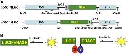
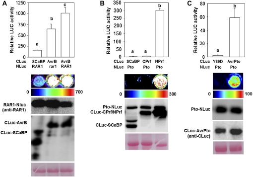
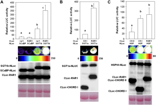
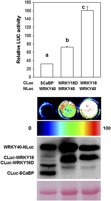
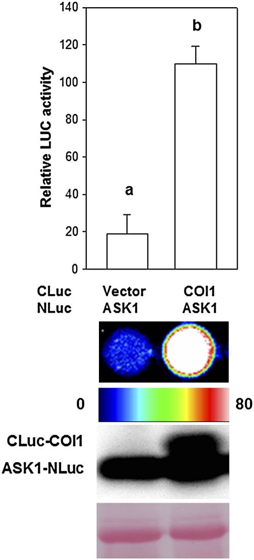
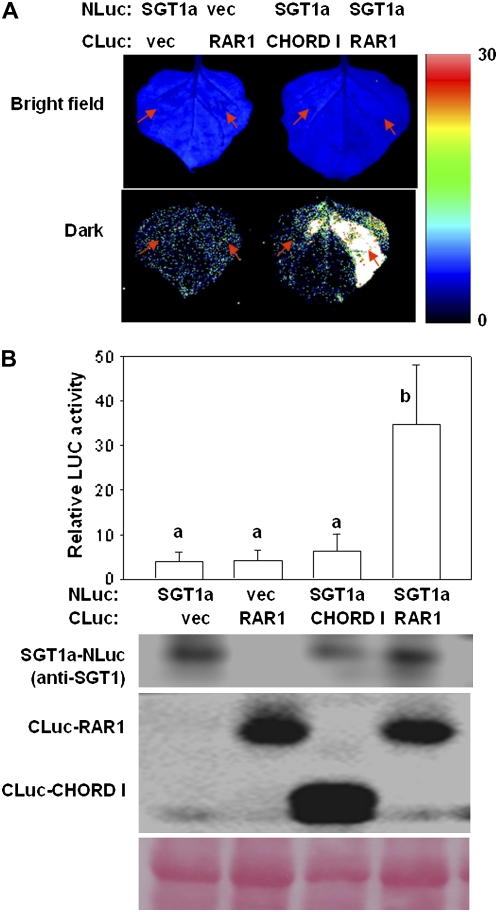
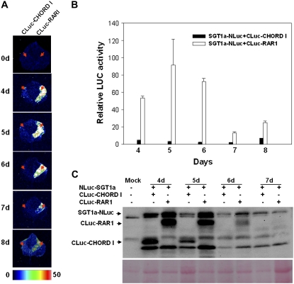
The development of sensitive and versatile techniques to detect protein-protein interactions in vivo is important for understanding protein functions. The previously described techniques, fluorescence resonance energy transfer and bimolecular fluorescence complementation, which are used widely for protein-protein interaction studies in plants, require extensive instrumentation. To facilitate protein-protein interaction studies in plants, we adopted the luciferase complementation imaging assay. The amino-terminal and carboxyl-terminal halves of the firefly luciferase reconstitute active luciferase enzyme only when fused to two interacting proteins, and that can be visualized with a low-light imaging system. A series of plasmid constructs were made to enable the transient expression of fusion proteins or generation of stable transgenic plants. We tested nine pairs of proteins known to interact in plants, including Pseudomonas syringae bacterial effector proteins and their protein targets in the plant, proteins of the SKP1-Cullin-F-box protein E3 ligase complex, the HSP90 chaperone complex, components of disease resistance protein complex, and transcription factors. In each case, strong luciferase complementation was observed for positive interactions. Mutants that are known to compromise protein-protein interactions showed little or much reduced luciferase activity. Thus, the assay is simple, reliable, and quantitative in detection of protein-protein interactions in plants.


Figures

Figure 1.
Figure 1.
Constructs for LCI assays in plants. A, Schematic diagrams of 35S∷NLuc and 35S∷CLuc constructs. L, Gly/Ser linker between LUC fragments and multiple cloning sites (MCS). rbs, Transcription terminator derived from the Rubisco small subunit gene. B, Diagram for LUC complementation resulting from NLuc- and CLuc-fusion proteins. [See online article for color version of this figure.]
Figure 2.
Figure 2.
Interactions of P. syringae effectors with host proteins in protoplasts. A, Interaction between AvrB and RAR1. B, Interaction between Pto and the N terminus of Prf. C, Interaction between AvrPto and Pto. The top panels show quantification of LUC activity. Different letters above the bars indicate statistic difference at P < 0.01 (t test). The images in the middle show microtiter plates containing protoplasts expressing the indicated constructs. The pseudocolor bar below shows the range of luminescence intensity in each image. The bottom panels show western blot for proteins isolated from protoplasts. Anti-full-length firefly LUC antibodies or the indicated specific antibodies (anti-RAR1; Shang et al., 2006; anti-CLuc antibodies; Sigma) were used to detect the indicated fusion proteins. The amount of protein loaded in each lane is indicated by Ponceau S staining of Rubisco on a representative protein blot. The data shown are representative of three independent experiments. [See online article for color version of this figure.]
Figure 3.
Figure 3.
Interactions among the HSP90 chaperone complex components in protoplasts. A, Interaction between SGT1b and RAR1. B, Interaction between SGT1a and RAR1. C, Interaction between RAR1 and HSP90. CH I, CHORD I domain; CH II, CHORD II domain; HSP, HSP90. The data shown are representative of three (A and B) or five (C) independent experiments. [See online article for color version of this figure.]
Figure 4.
Figure 4.
Interaction between WRKY40 and WRKY18. The WRKY18D construct lacks the Leu zipper motif. The data shown are representative of three independent experiments. [See online article for color version of this figure.]
Figure 5.
Figure 5.
Interactions between ASK1 and COI1. The data shown are representative of four independent experiments. [See online article for color version of this figure.]
Figure 6.
Figure 6.
Interactions between SGT1a and RAR1 in N. benthamiana leaves. A, LUC image of N. benthamiana leaves co-infiltrated with the agrobacterial strains containing SGT1a-NLuc and CLuc-RAR1. Arrows indicate leaf panels that were infiltrated with Agrobacterium containing the indicated constructs. B, Quantification of LUC activity in leaves expressing SGT1a-NLuc and CLuc-RAR1. The western blot below shows the expression levels of CLuc- and NLuc-fusion proteins. RAR1 derivatives were detected by anti-firefly LUC antibodies, whereas SGT1a-NLuc was detected by anti-SGT1 antibodies. Ponceau S staining shows equal loading of protein in lanes. Data were collected 4 d after infiltration. The data shown are representative of three independent experiments.
Figure 7.
Figure 7.
Time course of SGT1a-RAR1 interaction in N. benthamiana. A, LUC image of N. benthamiana leaves co-infiltrated with the agrobacterial strains containing SGT1a-NLuc and CLuc-RAR1. B, Quantification of LUC activity in leaves expressing SGT1a-NLuc and CLuc-RAR1. C, Accumulation of fusion proteins in N. benthamiana leaves. Data were collected at the indicated days post infiltration of Agrobacterium. Mock, Leaves infiltrated with water. Arrows indicate infiltrated regions. The data shown are representative of five independent experiments.

Similar articles

Cited by

References

    1. Azevedo C, Sadanandom A, Kitagawa K, Freialdenhoven A, Shirasu K, Schulze-Lefert P (2002) The RAR1 interactor SGT1, an essential component of R gene-triggered disease resistance. Science 295 2077–2080 - PubMed
    1. Bracha-Drori K, Shichrur K, Katz A, Oliva M, Angelovici R, Yalovsky S, Ohad N (2004) Detection of protein-protein interactions in plants using bimolecular fluorescence complementation. Plant J 40 419–427 - PubMed
    1. Bundock P, den Dulk-Ras A, Beijersbergen A, Hooykaas PJ (1995) Trans-kingdom T-DNA transfer from Agrobacterium tumefaciens to Saccharomyces cerevisiae. EMBO J 14 3206–3214 - PMC - PubMed
    1. Callis J, Vierstra RD (2000) Protein degradation in signaling. Curr Opin Plant Biol 3 381–386 - PubMed
    1. Chisholm ST, Coaker G, Day B, Staskawicz BJ (2006) Host-microbe interactions: shaping the evolution of the plant immune response. Cell 124 803–814 - PubMed

Publication types

MeSH terms