. 2008 Feb;74(3):633-44.
doi: 10.1128/AEM.02138-07. Epub 2007 Dec 7.

Characterization of htrB and msbB mutants of the light organ symbiont Vibrio fischeri

Affiliations

Characterization of htrB and msbB mutants of the light organ symbiont Vibrio fischeri

Dawn M Adin et al. Appl Environ Microbiol. 2008 Feb.

Abstract

Bacterial lipid A is an important mediator of bacterium-host interactions, and secondary acylations added by HtrB and MsbB can be critical for colonization and virulence in pathogenic infections. In contrast, Vibrio fischeri lipid A stimulates normal developmental processes in this bacterium's mutualistic host, Euprymna scolopes, although the importance of lipid A structure in this symbiosis is unknown. To further examine V. fischeri lipid A and its symbiotic function, we identified two paralogs of htrB (designated htrB1 and htrB2) and an msbB gene in V. fischeri ES114 and demonstrated that these genes encode lipid A secondary acyltransferases. htrB2 and msbB are found on the Vibrio "housekeeping" chromosome 1 and are conserved in other Vibrio species. Mutations in htrB2 and msbB did not impair symbiotic colonization but resulted in phenotypic alterations in culture, including reduced motility and increased luminescence. These mutations also affected sensitivity to sodium dodecyl sulfate, kanamycin, and polymyxin, consistent with changes in membrane permeability. Conversely, htrB1 is located on the smaller, more variable vibrio chromosome 2, and an htrB1 mutant was wild-type-like in culture but appeared attenuated in initiating the symbiosis and was outcompeted 2.7-fold during colonization when mixed with the parent. These data suggest that htrB2 and msbB play conserved general roles in vibrio biology, whereas htrB1 plays a more symbiosis-specific role in V. fischeri.


Figures

FIG. 1.
FIG. 1.
Chromosomal location and orientation of htrB1, htrB2, and msbB in V. fischeri ES114. Open reading frames encoding hypothetical proteins are designated “hyp.” Blast-P searches of the National Center for Biotechnology Information database were used to determine homologies (e < 10−15) and to assign names for genes flanking htrB1, htrB2, and msbB. Arrows indicate gene orientations. Ch1 and Ch2 indicate localization to chromosomes 1 and 2, respectively.
FIG. 2.
FIG. 2.
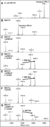
Complementation of E. coli MKV15 with putative lipid A secondary acyltransferase genes from V. fischeri. Lipid A was obtained from LPS preparations of E. coli strains grown in minimal medium at 30°C, including W3110 (parent) (A), MKV15 (mutant lacking lipid A secondary acylations) (B), and MKV15 carrying plasmids with either no insert (pDMA28) [C] or pJLB2 [F]) or inserts of htrB1, htrB2, and msbB from V. fischeri (D, E, and G, respectively). pDMA28 served as the isogenic insert-free control for pDMA25 and pDMA27, whereas pJLB2 served this purpose for pDMA114. Samples were analyzed by negative-ion MALDI-TOF MS in the reflectron mode. The (M − H) ion at m/z 1,796.1 (A) arises from E. coli DPLA. Strain MKV15 produces a tetra-acylated DPLA with an (M − H) ion at m/z 1,403.9 (B), which is also seen in MKV15 with the control plasmids (C and F). Arrows in panels D, E, and G indicate the molecular weight shifts caused by complementation. Peaks marked with asterisks are monophosphorylated lipid A forms. The small peak at m/z 1,622.0 (A and B) is an unidentified impurity. The minor addition of a C16:0 fatty acid (238 Da) occasionally occurred even in the negative controls (C, F, and G).
FIG. 3.
FIG. 3.
Motility of V. fischeri htrB1, htrB2, and msbB mutants. Both panels report average rates of movement through 0.25% agar plates and show representative data from one of at least experiments. Standard errors (n = 6) are shown. (A) Motility of ES114 and mutants. Bars labeled with the same capital letter do not differ significantly (P < 0.001), as determined by ANOVA. (B) Motility of ES114, DMA310 (htrB2), and HG320 (msbB) with pVSV105, pDMA43 (pVSV105 + htrB2), or pDMA42 (pVSV105 + msbB). Asterisks represent significant differences (P < 0.01) from the wild type carrying the same vector.
FIG. 4.
FIG. 4.
Morphology of htrB2 mutants. Scanning electron microscopy images of ES114 (wild type) (A), DMA310 (htrB2) (B), DMA330 (htrB1 htrB2) (C), and AO340 (htrB1 htrB2 msbB) (D) are shown.
FIG. 5.
FIG. 5.
Effects of htrB1, htrB2, and msbB mutations on bioluminescence. The maximal luminescence per OD595 for mutant strains grown in SWT medium at 24°C is expressed as a percentage of that emitted by ES114. Error bars indicate standard errors (n = 3), and asterisks indicate significant differences from ES114 (P < 0.05). Data for one representative experiment of three are shown.
FIG. 6.
FIG. 6.
Symbiotic luminescence and colonization of V. fischeri wild type and an htrB1 mutant. (A) Animal luminescence during the initial stages of E. scolopes colonization by strains ES114 (wild type) and EVS300 (htrB1 mutant). The luminescence pattern indicates the initial onset of colonization (0 to 12.5 h) followed by a changing level of light emission that reflects the diurnal venting behavior of the animal and regrowth of symbionts (2). Mean values for 21 animals were calculated, and standard errors of the means are indicated. (B) In a separate experiment, luminescence of hatchling squid inoculated with the wild type (ES114) or the htrB mutant (EVS300) was determined at 16 h postinoculation. Each bar represents the average for 11 or 12 animals, with the standard error. (C) Colonization levels of V. fischeri in the same animals as those presented in panel B. Each bar represents the average for 11 or 12 animals, with the standard error. Diamonds represent the CFU present in individual animals.
FIG. 7.
FIG. 7.
Competition between ES114 and htrB1 mutant EVS300 in culture and in squid. (A) Relative competitiveness of htrB1 cocultured with ES114 in LBS medium. In parallel cultures, one strain or the other was tagged with lacZ to facilitate determination of strain ratios by blue-white screening (see Materials and Methods). Error bars represent standard errors (n = 3). (B) Relative competitiveness of htrB1 mutant during colonization of E. scolopes. Juvenile squid were exposed to a mixed inoculum of the wild type and htrB1 mutant EVS300 at a total concentration of ∼2,500 CFU/ml, and the relative competitiveness was determined after 48 h. Circles represent the RCIs determined from infection of 118 individual animals. Circles with arrows represent animals that were clonally infected. Data represent combined results of four similar experiments.

References

    1. Boettcher, K. J., and E. G. Ruby. 1990. Depressed light emission by symbiotic Vibrio fischeri of the sepiolid squid Euprymna scolopes. J. Bacteriol. 172:3701-3706. - PMC - PubMed
    1. Boettcher, K. J., E. G. Ruby, and M. J. McFall-Ngai. 1996. Bioluminescence in the symbiotic squid Euprymna scolopes is controlled by daily biological rhythm. J. Comp. Physiol. A 179:65-73.
    1. Boylan, M., C. Miyamoto, L. Wall, A. Grahm, and E. Meighen. 1989. Lux C, D, and E genes of the Vibrio fischeri luminescence operon code for the reductase, transferase, and synthetase enzymes involved in aldehyde biosyntheses. Photochem. Photobiol. 49:681-688. - PubMed
    1. Caroff, M., L. Aussel, H. Zarrouk, A. Martin, J. C. Richards, H. Therisod, M. B. Perry, and D. Karibian. 2001. Structural variability and originality of the Bordetella endotoxins. J. Endotoxin Res. 7:63-68. - PubMed
    1. Chen, C. Y., K. M. Wu, Y. C. Chang, C. H. Chang, H. C. Tsai, T. L. Liao, Y. M. Liu, H. J. Chen, A. B. Shen, J. C. Li, T. L. Su, C. P. Shao, C. T. Lee, L. I. Hor, and S. F. Tsai. 2003. Comparative genome analysis of Vibrio vulnificus, a marine pathogen. Genome Res. 13:2577-2587. - PMC - PubMed

Publication types

LinkOut - more resources