

doi: 10.1038/nsmb1327.
Epub 2007 Dec 9.
Structural basis for the coevolution of a viral RNA-protein complex
Affiliations
- PMID: 18066080
- PMCID: PMC3152963
- DOI: 10.1038/nsmb1327
Item in Clipboard
Structural basis for the coevolution of a viral RNA-protein complex
Nat Struct Mol Biol.
2008 Jan.
Abstract
The cocrystal structure of the PP7 bacteriophage coat protein in complex with its translational operator identifies a distinct mode of sequence-specific RNA recognition when compared to the well-characterized MS2 coat protein-RNA complex. The structure reveals the molecular basis of the PP7 coat protein's ability to selectively bind its cognate RNA, and it demonstrates that the conserved beta-sheet surface is a flexible architecture that can evolve to recognize diverse RNA hairpins.
Figures
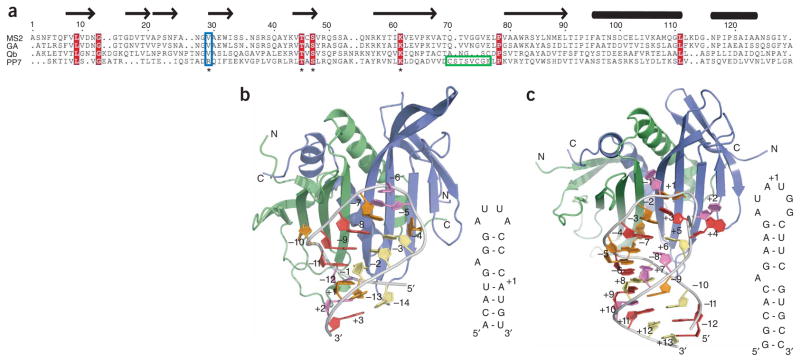
Coat-protein sequence alignment and overview of the MS2 coat protein and PP7ΔFG complexes with RNA. (a) Alignment of four ssRNA bacteriophage coat proteins. (b,c) MS2 coat protein–RNA (2BU1) (b) and PP7ΔFG–RNA (c) complexes are shown as cartoons. In both structures the RNA hairpin (orange, adenine; red, guanine; violet, uridine; yellow, cytidine) binds across the extended β-sheet surface formed by the coat protein dimer (blue, green).
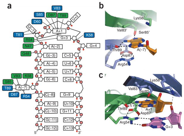
RNA-protein interface. (a) Schematic representation of PP7ΔFG interactions with the RNA hairpin. Black arrows, hydrogen bonds; black lines, van der Waals and stacking interactions. (b,c) The A(−7) (b) and A+1 (c) recognition pockets are shown, with the adenosine nucleotides (orange) and PP7ΔFG residues (blue, green) as sticks. Potential hydrogen bonds are shown as dashed lines.
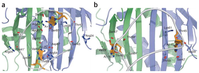
Orientation of adenine recognition pockets. (a,b) The position of the adenine recognition pockets is rotated approximately 90° with respect to the dimer axis between the PP7ΔFG (a) and MS2 (b) coat proteins. Cartoon ribbon shows the position of the phosphate backbone (gray) and bound adenosines are shown as sticks (orange).
References
Publication types
MeSH terms
Substances
Associated data
- Actions
- Actions
Grants and funding
LinkOut - more resources
Full Text Sources
Other Literature Sources
Molecular Biology Databases
Research Materials
