

Increased versican content is associated with tendinosis pathology in the patellar tendon of athletes with jumper's knee
- PMID: 18067512
- PMCID: PMC3951986
- DOI: 10.1111/j.1600-0838.2007.00735.x
Increased versican content is associated with tendinosis pathology in the patellar tendon of athletes with jumper's knee
Abstract
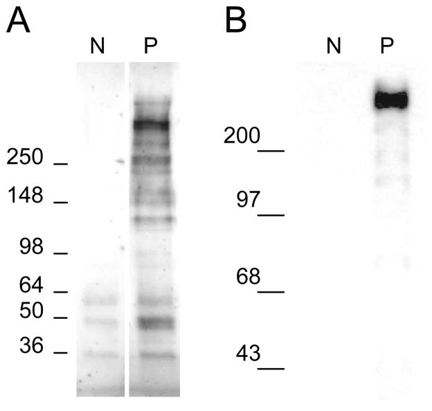
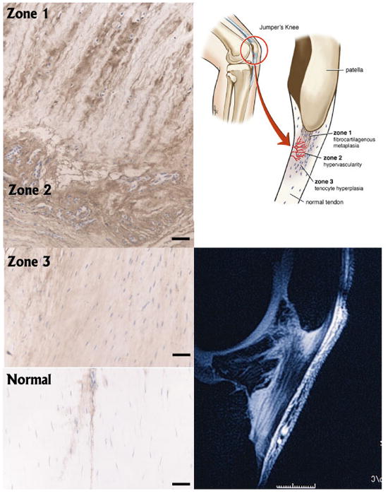
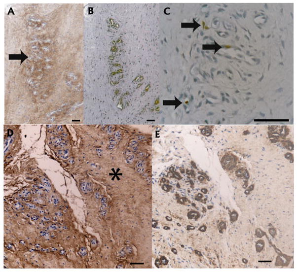
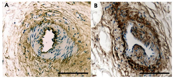
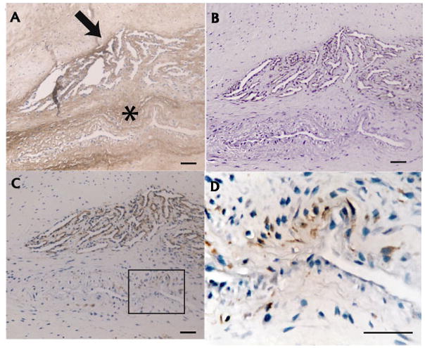
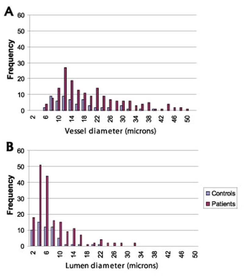
Expansion of the extracellular matrix is a prominent but poorly characterized feature of tendinosis. The present study aimed to characterize the extent and distribution of the large aggregating proteoglycan versican in patients with patellar tendinosis. We obtained tendon from tendinopathy patients undergoing debridement of the patellar tendon and from controls undergoing intramedullary tibial nailing. Versican content was investigated by Western blotting and immunohistochemistry. Microvessel thickness and density were determined using computer-assisted image analysis. Markers for smooth muscle actin, endothelial cells (CD31) and proliferating cells (Ki67) were examined immunohistochemically. Western blot analysis and immunohistochemical staining revealed elevated versican content in the proximal patellar tendon of tendinosis patients (P=0.042). Versican content was enriched in regions of fibrocartilage metaplasia and fibroblast proliferation, as well as in the perivascular matrix of proliferating microvessels and within the media and intima of arterioles. Microvessel density was higher in tendinosis tissue compared with control tissue. Versican deposition is a prominent feature of patellar tendinosis. Because this molecule is not only a component of normal fibrocartilagenous matrices but also implicated in a variety of soft tissue pathologies, future studies should further detail both pathological and adaptive roles of versican in tendons.
Figures
Similar articles
-
Changes in the composition of the extracellular matrix in patellar tendinopathy.Matrix Biol. 2009 May;28(4):230-6. doi: 10.1016/j.matbio.2009.04.001. Epub 2009 Apr 14. Matrix Biol. 2009. PMID: 19371780
-
Abnormal tenocyte morphology is more prevalent than collagen disruption in asymptomatic athletes' patellar tendons.J Orthop Res. 2004 Mar;22(2):334-8. doi: 10.1016/j.orthres.2003.08.005. J Orthop Res. 2004. PMID: 15013093
-
In vivo microdialysis and immunohistochemical analyses of tendon tissue demonstrated high amounts of free glutamate and glutamate NMDAR1 receptors, but no signs of inflammation, in Jumper's knee.J Orthop Res. 2001 Sep;19(5):881-6. doi: 10.1016/S0736-0266(01)00016-X. J Orthop Res. 2001. PMID: 11562137
-
Patellar tendinosis as an adaptive process: a new hypothesis.Br J Sports Med. 2004 Dec;38(6):758-61. doi: 10.1136/bjsm.2003.005157. Br J Sports Med. 2004. PMID: 15562176 Free PMC article. Review.
-
[Patellar tendinopathy ('jumper's knee'); a common and difficult-to-treat sports injury].Ned Tijdschr Geneeskd. 2008 Aug 16;152(33):1831-7. Ned Tijdschr Geneeskd. 2008. PMID: 18783161 Review. Dutch.
Cited by
-
Regenerative biology of tendon: mechanisms for renewal and repair.Curr Mol Biol Rep. 2015 Sep;1(3):124-131. doi: 10.1007/s40610-015-0021-3. Curr Mol Biol Rep. 2015. PMID: 26389023 Free PMC article.
-
A New Quantitative Tool for the Ultrasonographic Assessment of Tendons: A Reliability and Validity Study on the Patellar Tendon.Diagnostics (Basel). 2024 May 21;14(11):1067. doi: 10.3390/diagnostics14111067. Diagnostics (Basel). 2024. PMID: 38893594 Free PMC article.
-
The "other" 15-40%: The Role of Non-Collagenous Extracellular Matrix Proteins and Minor Collagens in Tendon.J Orthop Res. 2020 Jan;38(1):23-35. doi: 10.1002/jor.24440. Epub 2019 Aug 26. J Orthop Res. 2020. PMID: 31410892 Free PMC article. Review.
-
Overload and neovascularization of shoulder tendons in volleyball players.BMC Res Notes. 2012 Aug 1;5:397. doi: 10.1186/1756-0500-5-397. BMC Res Notes. 2012. PMID: 22853746 Free PMC article.
-
Proteomic differences between male and female anterior cruciate ligament and patellar tendon.PLoS One. 2014 May 12;9(5):e96526. doi: 10.1371/journal.pone.0096526. eCollection 2014. PLoS One. 2014. PMID: 24818782 Free PMC article.
References
-
- Almekinders LC, Weinhold PS, Maffulli N. Compression etiology in tendinopathy. Clin Sports Med. 2003;22:703–10. - PubMed
-
- Archambault JM, Jelinsky SA, Lake SP, Hill AA, Glaser DL, Soslowsky LJ. Rat supraspinatus tendon expresses cartilage markers with overuse. J Orthop Res. 2007;25:617–24. - PubMed
-
- Banes AJ, Horesovsky G, Larson C, Tsuzaki M, Judex S, Archambault J, Zernicke R, Herzog W, Kelley S, Miller L. Mechanical load stimulates expression of novel genes in vivo and in vitro in avian flexor tendon cells. Osteoarthritis Cartilage. 1999;7:141–53. - PubMed
-
- Bernstein EF, Fisher LW, Li K, LeBaron RG, Tan EM, Uitto J. Differential expression of the versican and decorin genes in photoaged and sun-protected skin. Comparison by immunohistochemical and northern analyses. Lab Invest. 1995;72:662–9. - PubMed
-
- Biberthaler P, Wiedemann E, Nerlich A, Kettler M, Mussack T, Deckelmann S, Mutschler W. Microcirculation associated with degenerative rotator cuff lesions. In vivo assessment with orthogonal polarization spectral imaging during arthroscopy of the shoulder. J Bone Joint Surg Am. 2003;85-A:475–80. - PubMed
Publication types
MeSH terms
Substances
Grants and funding
LinkOut - more resources
Full Text Sources
Medical
