. 2008 Jan;67(2):435-47.
doi: 10.1111/j.1365-2958.2007.06057.x. Epub 2007 Dec 5.

Genomic rearrangements and transcriptional analysis of the spliced leader-associated retrotransposon in RNA interference-deficient Trypanosoma brucei

Affiliations

Genomic rearrangements and transcriptional analysis of the spliced leader-associated retrotransposon in RNA interference-deficient Trypanosoma brucei

Kristin L Patrick et al. Mol Microbiol. 2008 Jan.

Abstract

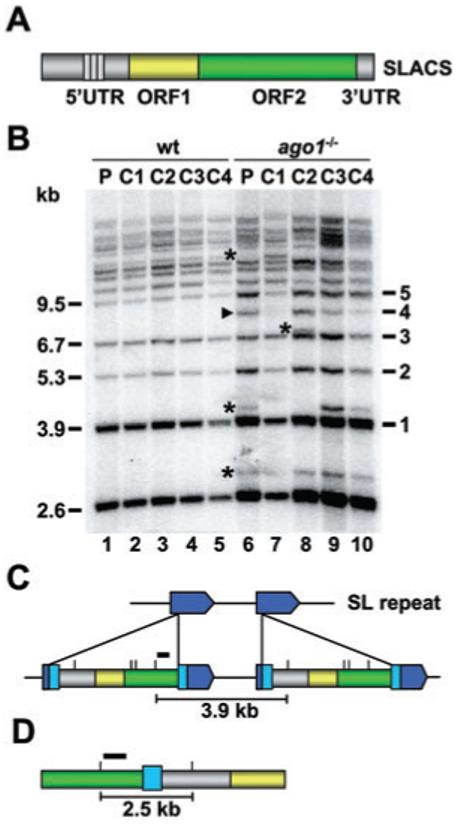
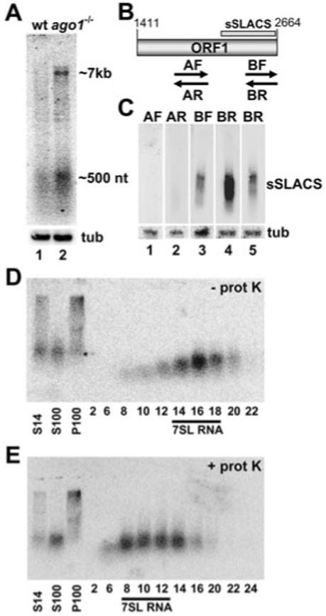
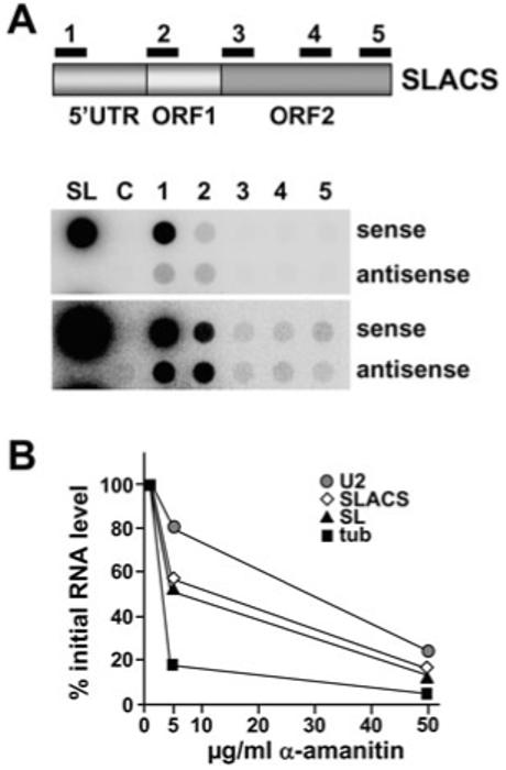
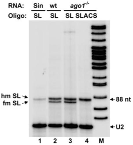
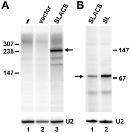
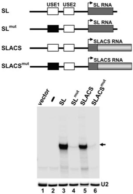
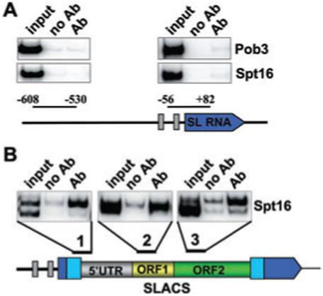
The Trypanosoma brucei genome is colonized by the site-specific non-LTR retrotransposon SLACS, or spliced leader-associated conserved sequence, which integrates exclusively into the spliced leader (SL) RNA genes. Although there is evidence that the RNA interference (RNAi) machinery regulates SLACS transcript levels, we do not know whether RNAi deficiency affects the genomic stability of SLACS, nor do we understand the mechanism of SLACS transcription. Here, we report that prolonged culturing of RNAi-deficient T. brucei cells, but not wild-type cells, results in genomic rearrangements of SLACS. Furthermore, two populations of SLACS transcripts persist in RNAi-deficient cells: a full-length transcript of approximately 7 kb and a heterogeneous population of small SLACS transcripts ranging in size from 450 to 550 nt. We provide evidence that SLACS transcription initiates at the +1 of the interrupted SL RNA gene and proceeds into the 5' UTR and open reading frame 1 (ORF1). This transcription is carried out by an RNA polymerase with alpha-amanitin sensitivity reminiscent of SL RNA synthesis and is dependent on the SL RNA promoter. Additionally, we show that both sense and antisense small SLACS transcripts originate from ORF1 and that they are associated with proteins in vivo. We speculate that the small SLACS transcripts serve as substrates for the production of siRNAs to regulate SLACS expression.


Figures

Fig. 1
Fig. 1
Genomic organization of SLACS in the SL RNA gene cluster of T. brucei rhodesiense. A. Schematic representation of the SLACS element with the 5′ UTR, ORF1, ORF2 and 3′ UTR indicated in different colours. The 185 bp repeat region present in the 5′ UTR is shown by small rectangles. The poly(dA) tract immediately following the 3′ UTR has been omitted for clarity. B. Southern blot analysis of wild-type (wt) and ago1-/- genomic DNA digested with EcoRV and hybridized with a probe indicated by a black bar in panels C and D. The wild-type (P, lane 1) and ago1-/- populations (P, lane 6) were cloned, and four randomly selected clones were analysed for both cell types (C1-C4, lanes 2-5 and lanes 7-10). Black arrow heads and asterisks indicate new bands appearing in ago1-/- cells after being maintained in culture for 8 months. Approximate sizes (kb) and the number of SL RNA repeats separating SLACS elements are indicated on the left and right respectively. C. Schematic representation of two SLACS elements integrated into two adjacent SL RNA genes, shown in dark blue. The 49 bp duplication of the target sequence, generated as a consequence of SLACS insertion, is shown in light blue. Small vertical lines above the SLACS elements specify EcoRV restriction sites and the black bar identifies the probe used for the Southern blot analysis shown in panel B. And 3.9 kb is the predicted distance between EcoRV sites in two adjacent SLACS. D. Schematic representation of the cloned 2.5 kb genomic fragment, with two tandem SLACS separated by the 49 bp target site duplication. The black bar identifies the probe used for Southern blot analysis shown in panel D. Drawings are not to scale.
Fig. 2
Fig. 2
Small SLACS transcripts in ago1-/- cells. A. Northern blot analysis of SLACS transcripts in wild-type (wt, lane 1) and ago1-/- cells (lane 2) using a probe designed against SLACS ORF1. Approximate sizes of full-length SLACS transcripts (∼7 kb) and the sSLACS transcripts (∼500 nt) are indicated. α-Tubulin (tub) served as a control for RNA recovery and loading (lower panel). B. Schematic representation of SLACS ORF1. The origin of the sSLACS is indicated by a bar and the PCR-synthesized single-stranded probes used in panel C are shown below ORF1. The drawing is to scale. C. Northern blot analysis of sSLACS transcripts. RNA from ago1-/- cells was hybridized with probe AF (lane 1) and AR (lane 2), which detect antisense and sense transcripts respectively, between SLACS nt 1755-2054 (see panel B), and with probes BF (lane 3) and BR (lane 4), which detect antisense and sense transcripts respectively, between SLACS nt 2359-2661 (see panel B). A shorter exposure of the BR blot is provided at the far right. α-Tubulin (tub) served as a control for RNA recovery and loading (lower panel). D. A post-nuclear supernatant from ago1-/- cells (S14) was separated by high-speed centrifugation in a soluble (S100) and pellet fraction (P100), and the S100 was applied to a 10-30% glycerol density gradient. Selected fractions were Northern-blotted with a probe complementary to SLACS ORF1. The fractions containing 7SL RNA are indicated. E. An equivalent amount of the S100 fraction that was analysed in D was digested with proteinase-K prior to glycerol density gradient centrifugation. The fractions containing 7SL RNA are indicated.
Fig. 3
Fig. 3
SLACS transcription in ago1-/- cells. A. Dot blot hybridization of newly synthesized RNA DNA fragments of about 300 bp, cloned into M13 vectors and spotted onto a nitrocellulose membrane. The numbers refer to the regions indicated in the accompanying graphical representation of SLACS. Drawing is not to scale. SL, spliced leader RNA gene probe; C, vector control. Two different exposure times are shown. B. α-Amanitin sensitivity of SLACS transcription. [α-32P]-RNA was synthesized in permeable cells in the presence of increasing concentrations of α-amanitin as indicated, hybridized to cloned DNAs immobilized onto nitrocellulose filters, quantified and expressed as percentages of the level of transcription measured in the absence of the inhibitor. U2, U2 snRNA; tub, α-tubulin.
Fig. 4
Fig. 4
SLACS transcripts initiate at the +1 nt of the interrupted SL RNA gene. Total RNA isolated from sinefungin-treated (sin, lane 1), wild-type (wt, lane 2) and ago1-/- cells (lanes 3 and 4) was primer-extended with an oligonucleotide complementary to nt 65-88 of the SL RNA (SL, lanes 1-3) and an oligonucleotide complementary to SLACS nt 1-28 (SLACS, lane 4). Primer extension stops corresponding to fully modified (fm) and hypomodified (hm) SL RNA are indicated. M, α-32P-labelled MspI digest of pBR322. A U2 snRNA-specific primer was included in all the reactions to control for RNA amounts and quality (U2).
Fig. 5
Fig. 5
SLACS in vitro transcription. A. In vitro transcription reactions containing no added DNA (lane 1), the pXS2 backbone vector (lane 2) or pSLACS (lane 3) were performed in the presence of [α-32P]-GTP, and RNA products were analysed on an 8% polyacrylamide-7 M urea gel. B. In vitro transcription reactions were carried out in the absence of radiolabelled nucleotides, and RNA was analysed by primer extension with an end-labelled oligonucleotide complementary to a 19 nt tag engineered in both the SLACS and SL templates (SLtag). U2 primer extension served as a loading control in both panels.
Fig. 6
Fig. 6
SLACS transcription is dependent on the upstream SL promoter. In vitro transcription reactions were carried out with the constructs schematically shown above the autoradiograph, and RNA was primer-extended with the SLtag oligonucleotide as described in Fig. 5. Drawings are not to scale. U2 primer extension served as a loading control (U2).
Fig. 7
Fig. 7
Spt16 association with SLACS, but not the SL RNA gene. ChIP was performed with antibodies directed against the BB2 epitope present on Spt16 or Pob3, and precipitates were analysed by PCR with primers specific for the regions indicated (Ab). A control with no antibody (no Ab) and an aliquot of the total input (input) is also shown. The double bands observed at SLACS regions 1 and 3 are a consequence of sequence heterogeneity, i.e. nucleotide deletions/insertions, in the 5′ UTR and ORF2, respectively, of SLACS elements.

References

    1. Aksoy S, Lalor TM, Martin J, Van der Ploeg LH, Richards FF. Multiple copies of a retroposon interrupt spliced leader RNA genes in the African trypanosome, Trypanosoma gambiense. EMBO J. 1987;6:3819–3826. - PMC - PubMed
    1. Aksoy S, Williams S, Chang S, Richards FF. SLACS retrotransposon from Trypanosoma brucei gambiense is similar to mammalian LINEs. Nucleic Acids Res. 1990;18:785–792. - PMC - PubMed
    1. Aravin AA, Naumova NM, Tulin AV, Vagin VV, Rozovsky YM, Gvozdev VA. Double-stranded RNA-mediated silencing of genomic tandem repeats and transposable elements in the D. melanogaster germline. Curr Biol. 2001;11:1017–1027. - PubMed
    1. Axtell MJ, Jan C, Rajagopalan R, Bartel DP. A two-hit trigger for siRNA biogenesis in plants. Cell. 2006;127:565–577. - PubMed
    1. Baulcombe DC. Molecular biology. Amplified silencing. Science. 2007;315:199–200. - PubMed

Publication types