. 2008 May;124(1):129-40.
doi: 10.1111/j.1365-2567.2007.02749.x. Epub 2007 Dec 7.

CD40 Ligand-activated, antigen-specific B cells are comparable to mature dendritic cells in presenting protein antigens and major histocompatibility complex class I- and class II-binding peptides

Affiliations

CD40 Ligand-activated, antigen-specific B cells are comparable to mature dendritic cells in presenting protein antigens and major histocompatibility complex class I- and class II-binding peptides

Tahamtan Ahmadi et al. Immunology. 2008 May.

Abstract

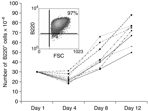
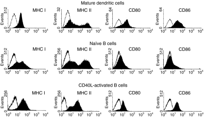
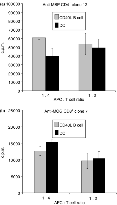
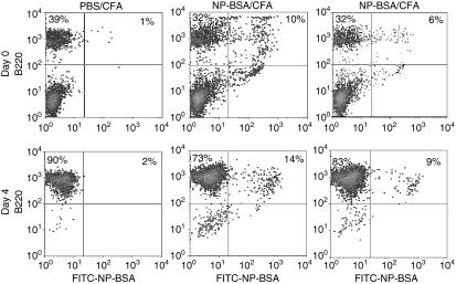
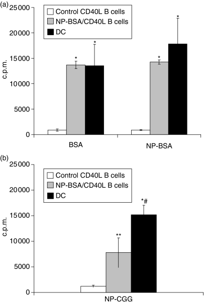
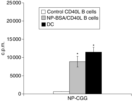
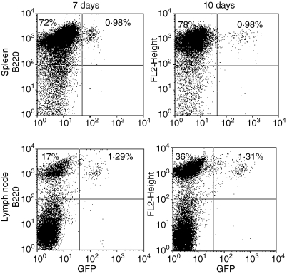
Dendritic cells (DC) are increasingly exploited for cell-based immunotherapy. However, limitations in ex vivo DC growth and DC functional heterogeneity have motivated development of complementary antigen-presenting cell sources. Here, the ability of CD40 ligand (CD40L)-activated B cells to fulfil that role was investigated. We demonstrate for the first time that non-specific or antigen-specific murine B cells can be grown for extended periods of time by stimulation with CD40L. These cells rapidly up-regulate and maintain high levels of co-stimulatory molecules. In a head-to-head comparison with DC, CD40L-expanded B cells were comparable to DC in the presentation of peptides to CD4+ and CD8+ T cells. While DC were superior to antigen non-specific CD40L-activated B cells with regard to whole protein (NP-BSA) processing and presentation, CD40L-expanded B cells from NP-BSA-immunized mice were comparable to DC when presenting BSA or NP-BSA to primed primary T cells or when presenting NP linked to an unrelated carrier, CGG, to naïve T cells. Thus, the combination of CD40L activation, which supports B-cell growth and augments intracellular protein processing, and antigen uptake via the B-cell receptor, allows for efficient uptake, processing, and presentation of whole protein antigens in a fashion comparable to that observed with mature DC. Like DC, CD40L-activated B cells efficiently home to secondary lymphoid organs in vivo. This system represents a unique tool for studying primary antigen-specific B cells and the results suggest that the outgrowth of large numbers of highly activated B cells represents a viable and practical complement to DC for cell-based immunotherapy.


Figures

Figure 1
Figure 1
Murine B-cell populations can be expanded by stimulation with CD40L. Spleen cell populations were phenotyped for B220 expression and cultured in duplicate wells for 12 days on CD40L-transfected L cells in the presence of rIL-4. Duplicate wells were harvested on days 4, 8, and 12 and the number of viable (trypan blue excluding), B220+ B cells determined. Data represent the total number of B220+ B cells recovered at various time points in 8 independent cultures. A representative B220 phenotype of cells after 7 days of culture is presented in the insert.
Figure 2
Figure 2
Murine CD40L-activated B cells exhibit a similar co-regulatory molecule phenotype as dendritic cells. Mature DC were generated from murine bone marrow by culture with rIL-4 and rGM-CSF for 4 days followed by a two day culture with rTNF-α. CD40L-activated B cells were generated by culture for 12 days on monolayers of L cells transfected with human CD40L in the presence of rIL-4 and cyclosporin A. Cells were phenotyped using a combination of B220-specific antibody and antibody specific for MHC I, MHC II, CD80, or CD86. Co-regulatory molecule expression (solid histograms) on cells gated for B220 expression is presented. Open histograms represent data generated with FITC-isotype control antibodies. Data from a representative experiment (>5 total) are presented.
Figure 3
Figure 3
Murine CD40L-activated B cells are comparable to mature dendritic cells (DC) in peptide presentation to CD4+ and CD8+ T-cell clones. Bone marrow DC or splenic CD40L-activated and expanded B-cell populations were generated as in Fig. 1 and loaded with 10 μg/ml MOG or MBP peptides, irradiated and cultured for 36 hr with MBP/MHC II-restricted CD4+ T cells (‘clone 12’) or MOG/MHC I-restricted, CD8+ T cells (‘clone 7’). 3H-thymidine was added and CPM incorporation determined 18–20 hr later. Data obtained from 3 independent cultures of APC are presented as means (3H-thymidine incorporation in the presence of peptide – 3H-thymidine incorporation in the absence of peptide) ± SE. Background incorporation of CD8+ and CD4+ T-cell clones cultured in the presence of APC but without peptide averaged <600 c.p.m. and <2000 c.p.m., respectively.
Figure 4
Figure 4
Growth of antigen-specific B cells after CD40L activation. Mice were injected three times with PBS/CFA or NP-BSA/CFA. Ten days after the last treatment, mice were killed and an aliquot of spleen cells stained with PE–anti-B220 antibody and with FITC–conjugated NP-BSA. The remainder of the spleen cells were cultured on CD40L-transfected L cells in the presence of rIL-4 and cyclosporin A for 4 days and phenotyped. Representative data from a total of 9 mice on day 0 and 6 mice on day 4 are presented.
Figure 5
Figure 5
Antigen-specific CD40L-activated B cells efficiently endocytose and present protein antigen to primed primary T cells. Mice were injected with PBS/CFA or NP-BSA/CFA as in Fig. 4. Ten days after the last treatment, mice were killed and splenocytes cultured for a minimum of 10 days on CD40L-transfected L cells in the presence of rIL-4. CD40L-activated B cells from PBS/CFA-treated (control) mice, CD40L-activated B cells from NP-BSA/CFA-immunized mice, or DC from control mice were pulsed with 1 μg/ml BSA or NP-BSA (a), or NP-CGG (b), irradiated, and 5 × 10⁴ cells added to 105 purified T cells from NP-BSA-immunized mice. After 36 hr, 3H-thymidine was added and c.p.m. incorporation determined 18–20 hr thereafter. Data are presented as means from three independent cultures of each APC (3H-thymidine incorporation in the presence of peptide – 3H-thymidine incorporation in the absence of peptide) ± SE. Background incorporation of T cells cultured in the presence of APC but without peptide averaged <600 c.p.m. and <2800 c.p.m., respectively. Significant differences relative to responses generated in the presence of antigen-pulsed, control CD40L-activated B cells, *P < 0·003 and **P < 0·02, respectively. Significant difference relative to responses generated in the presence NP-CGG-pulsed CD40L-activated B cells from NP-BSA/CFA-immunized mice, #P < 0·03.
Figure 6
Figure 6
Hapten-specific CD40L-activated B cells are comparable to DC in their ability to present hapten–protein to antigen naive, primary T cells. CD40L-activated B cells and DCs were pulsed with 1 μg/ml NP-CGG, washed extensively, irradiated, and cultured for 36 hr with T cells from PBS/CFA-treated mice. 3H-Thymidine was added and c.p.m. incorporation determined 18–20 hr thereafter. Data are presented as means from three independent cultures of each APC (3H-thymidine incorporation in the presence of peptide –3H-thymidine incorporation in the absence of peptide) ± SE. Significant difference relative to responses generated with NP-CGG pulsed CD40L-activated B cells expanded from control mice, *P < 0·005.
Figure 7
Figure 7
CD40L-activated B cells efficiently home to secondary lymphoid organs. Single-cell suspensions from GFP transgenic mice were cultured on CD40L-transfected L cells in the presence of rIL-4 and cyclosporin as in the legend to Fig. 1. Seven days later cells were washed and 3 × 10⁷ cells injected i.v. into C57BL/6 mice (four mice/group). Mice were killed 7 or 10 days later. Spleens and inguinal lymph nodes were removed, stained with PE-labelled B220-specific or isotype-control antibody and analysed by flow cytometry. Quadrants were set using PE–isotype antibody-treated C57BL/6 spleen or lymph node cells. Data presented were gated on lymphocytes, as determined by forward and side scatter parameters, and are representative of results obtained in each of four mice killed 7 and 10 days after cell injection. The percentage of cells falling into the B220+/GFP and B220+/GFP+ upper left and upper right quadrants respectively are indicated.

Similar articles

Cited by

References

    1. Steinman RM, Cohn ZA. Identification of a novel cell type in peripheral lymphoid organs of mice. I. Morphology, quantitation, tissue distribution. J Exp Med. 1973;137:1142–62. - PMC - PubMed
    1. Nestle FO, Alijagic S, Gilliet M, et al. Vaccination of melanoma patients with peptide- or tumor lysate-pulsed dendritic cells. Nat Med. 1998;4:328–32. - PubMed
    1. Brossart P, Wirths S, Stuhler G, Reichardt VL, Kanz L, Brugger W. Induction of cytotoxic T-lymphocyte responses in vivo after vaccinations with peptide-pulsed dendritic cells. Blood. 2000;96:3102–8. - PubMed
    1. Banchereau J, Schuler-Thurner B, Palucka AK, Schuler G. Dendritic cells as vectors for therapy. Cell. 2001;106:271–4. - PubMed
    1. Banchereau J, Palucka AK, Dhodapkar M, et al. Immune and clinical responses in patients with metastatic melanoma to CD34(+) progenitor-derived dendritic cell vaccine. Cancer Res. 2001;61:6451–8. - PubMed

Publication types

MeSH terms