Review
. 2008;110(3):182-95.
doi: 10.1016/j.acthis.2007.10.003. Epub 2007 Dec 18.

Glial cells: old cells with new twists

Affiliations
Review

Glial cells: old cells with new twists

Ugo Ndubaku et al. Acta Histochem. 2008.

Abstract

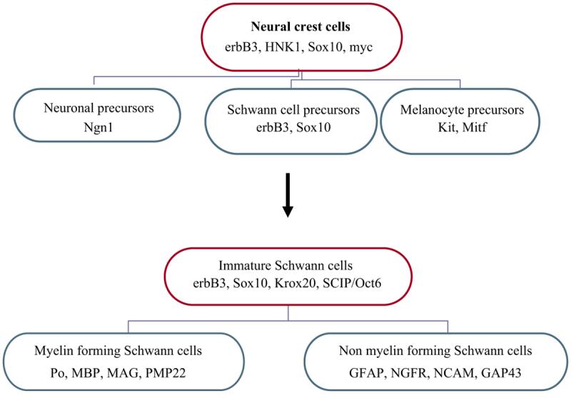
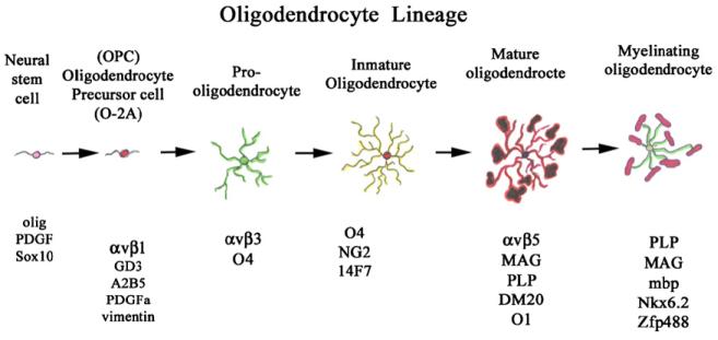
Based on their characteristics and function--migration, neural protection, proliferation, axonal guidance and trophic effects--glial cells may be regarded as probably the most versatile cells in our body. For many years, these cells were considered as simply support cells for neurons. Recently, it has been shown that they are more versatile than previously believed--as true stem cells in the nervous system--and are important players in neural function and development. There are several glial cell types in the nervous system: the two most abundant are oligodendrocytes in the central nervous system and Schwann cells in the peripheral nervous system. Although both of these cells are responsible for myelination, their developmental origins are quite different. Oligodendrocytes originate from small niche populations from different regions of the central nervous system, while Schwann cells develop from a stem cell population (the neural crest) that gives rise to many cell derivatives besides glia and which is a highly migratory group of cells.


Figures

Figure 1
Figure 1
Schwann cell development. The diagram shows how Schwann cells develop from a pluripotent neural crest cell that can give rise to different cells, from which the immature and mature Schwann cells will originate. Mature Schwann cells can be either myelin or nonmyelinating type. Ngn1 (neurogenin-1), Mitf (microphthalmia transcription factor), myc (myelocytomatosis viral oncogene), GFAP (glial fibrillary protein), NGFR (nerve growth factor receptor), NCAM (Neural cell adhesion protein), GAP43 (growth-associated protein 43), Krox20, SCIP/Oct6 (Octamer-binding transcription factor 6).
Figure 2
Figure 2
Schwann cell precursor during chicken development. Schwann cell precursors (SCP) were visualized by in situ hybridization using a probe specific to Seraf. SCP can be observed from the very early stages of neural crest development (arrows in A–C). At later stages of development (C, D), when mature Schwann cells are present, Seraf still labels these cells along forming nerves (arrowheads in C point to spinal nerves and in D points to hindlimb nerve plexus). Notice the distinct Seraf pattern in Figure 3C; arrows show migrating neural crest cells positive for Seraf, while arrowheads show staining of spinal nerve). HH = Hamburger and Hamilton developmental stages.
Figure 3
Figure 3
A summary of the best-known markers for the different stages of development of the oligodendrocyte. Not all researchers agree on the arbitrary stages of development shown here, but this combined figure highlights most of the current research on their development. OPC (oligodendrocyte precursor cell); O-2A (oligodendrocyte-astrocyte type 2A precursor); Olig (oligodendrocyte marker-1 and 2); PDGF (platelet derived growth factor); αvβ1 (integrin alpha v and beta-1 subunits); GD3 (ganglioside 3); A2B5; αvβ3; O4 (oligodendrocyte marker-4); NG2 (chondroitin sulfate proteoglycan); 14F7 (for an antibody with that nomenclature); αvβ5; MAG (myelin-associated glycoprotein); PLP/DM20 (proteolipid protein); O1 (oligodendrocyte marker-1); MBP (myelin basic protein).

References

    1. Allinquant B, Staugaitis SM, D"Urso D, Colman DR. The ectopic expression of myelin basic protein isoforms in Shiverer oligodendrocytes: implications for myelino-genesis. J Cell Biol. 1991;113:393–403. - PMC - PubMed
    1. Appel B, Givan LA, Eisen JS. Delta-notch signaling and lateral inhibition in zebrafish spinal cord development. BMC Dev Biol. 2001;1:13. - PMC - PubMed
    1. Asakura K, Miller DJ, Murray K, Bansal R, Pfeiffer SE, Rodriguez M. Monoclonal autoantibody SCH94.03, which promotes central nervous system remyelination, recognizes an antigen on the surface of oligodendrocytes. J Neurosci Res. 1996;43:273–81. - PubMed
    1. Augustine K, Liu ET, Sadler TW. Antisense attenuation of Wnt-1 and Wnt-3a expression in whole embryo culture reveals roles for these genes in craniofacial, spinal cord, and cardiac morphogenesis. Dev Genet. 1993;14:500–20. - PubMed
    1. Baker CV, Bronner-Fraser M. The origins of the neural crest. Part II: An evolutionary perspective. Mech Dev. 1997;69:13–29. - PubMed

LinkOut - more resources