. 2007 Dec;12(6):572-85.
doi: 10.1016/j.ccr.2007.11.002.

Enhanced paracrine FGF10 expression promotes formation of multifocal prostate adenocarcinoma and an increase in epithelial androgen receptor

Affiliations

Enhanced paracrine FGF10 expression promotes formation of multifocal prostate adenocarcinoma and an increase in epithelial androgen receptor

Sanaz Memarzadeh et al. Cancer Cell. 2007 Dec.

Abstract

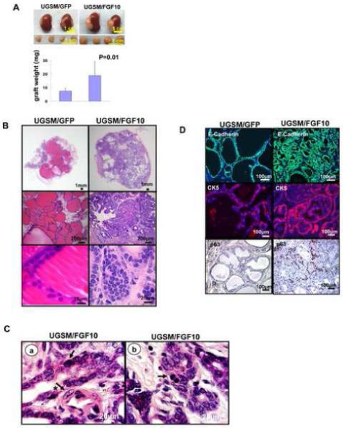
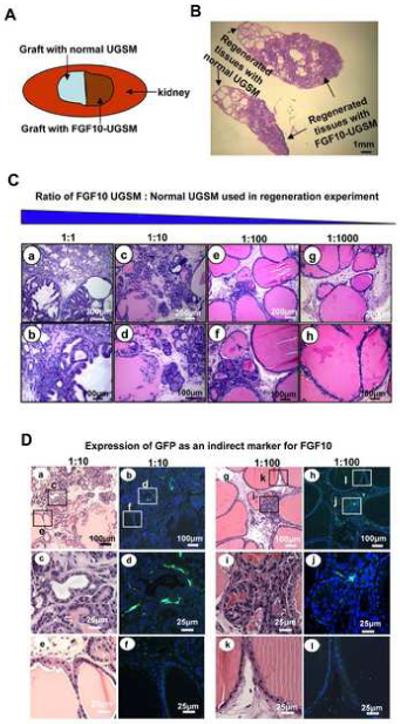
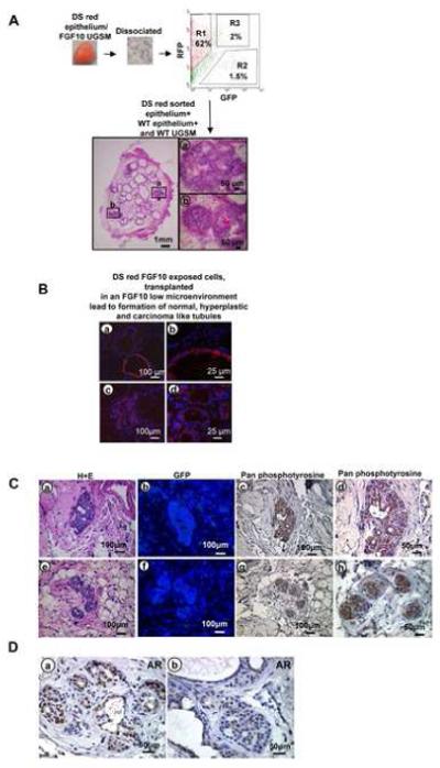
Enhanced mesenchymal expression of FGF10 led to the formation of multifocal PIN or prostate cancer. Inhibition of epithelial FGFR1 signaling using DN FGFR1 led to reversal of the cancer phenotype. A subset of the FGF10-induced carcinoma was serially transplantable. Paracrine FGF10 led to an increase in epithelial androgen receptor and synergized with cell-autonomous activated AKT. Our observations indicate that stromal FGF10 expression may facilitate the multifocal histology observed in prostate adenocarcinoma and suggest the FGF10/FGFR1 axis as a potential therapeutic target in treating hormone-sensitive or refractory prostate cancer. We also show that transient exposure to a paracrine growth factor may be sufficient for the initiation of oncogenic transformation.


Figures

Figure1
Figure1
Paracrine FGF10 expression led to well-differentiated adenocarcinoma and an expansion of both luminal and basal cells. A: Transilluminating image and weight (mean±SD) of regenerated tissue from wild type epithelia (1×10⁵) combined with GFP or FGF10 expressing mesenchyme (1×10⁵) placed in regeneration. B: H&E analysis of FGF10 UGSM and GFP UGSM regenerated tissue. C: High power (1000x) magnification of FGF10-induced adenocarcinoma demonstrating atypical (arrow), large nuclei (arrow and asterisk) in a and evidence of mitotic figures (arrow) in b. D: Immunohistochemical analysis of the regenerated tissue using antibodies against E-cadherin, CK 5 and P63.
Figure 2
Figure 2
Paracrine FGF10 signaling promoted formation of multifocal prostate cancer resembling human PIN or prostate cancer, and exerted its effects with regional specificity. A: Schematic representation of two adjacent grafts, one with FGF10 UGSM (1×10⁵) and a second graft with normal UGSM (1×10⁵) both combined with WT epithelium. B: Histology of two adjacent grafts revealed normal prostate tubules on one side and well-differentiated prostate cancer on the other side, suggesting localized action of paracrine FGF10. C: H&E analysis of regenerated tissue after a logarithmic reduction in the number of FGF10 UGSM revealed evidence of multifocal carcinoma. Cancer was seen in a&b, PIN adjacent to microinvasive carcinoma in c&d, multifocal PIN in e&f and predominantly normal histology was seen in g&h. D: Expression of FGF10 indirectly detected by anti-GFP staining in areas of hyperplasia.
Figure 3
Figure 3
FGF10 induces prostate cancer predominantly by activation of FGFR1. A. The lentivirus FUCRW vector used for the expression of flag tagged DN FGFR1 and DN FGF R2. Western blot confirmed the expression of both dominant negative constructs. B: Comparative analysis of graft weights (mean±SD) regenerated with control vector RFP, DN FGFR1-RFP or DN FGFR2-RFP and combined with FGF10 (5×10⁴) added to WT (5×10⁴) UGSM. C: Immunofluorescent analysis of grafts regenerated with vector control RFP, DN FGFR1-RFP, or DN FGFR2-RFP. DN FGFR1-RFP infected tubules showed cells completely confined within simple glandular and tubular structures suggestive of normal prostate epithelium in contrast to RFP or DN FGFR2-RFP regenerated glands.
Figure 4
Figure 4
Paracrine FGF10 resulted in an increase in epithelial AR A: Western blot revealed a four-fold increase in expression of AR protein in FGF10 compared to control regenerated grafts. B: Immunohistochemistry confirmed that paracrine FGF10 induced an increase in glandular epithelial AR compared to control regenerated tissue.
Figure 5
Figure 5
Paracrine FGF10 promoted androgen independent survival of a subset of prostate adenocarcinoma A: Histology of the FGF10 and WT mesenchyme regenerated grafts harvested from castrated animals. Contrary to normal WT regenerated grafts, persistence of adenocarcinoma was noted in all five FGF10 regenerated specimens. B: Immunohistochemical localization of AR in FGF10 grafts obtained from intact and castrated animals. Castration led to partial cytoplasmic transport of AR in castrate (c&d) compared to intact grafts (a&b). Partial nuclear localization of AR persisted despite castration in the FGF10 regenerated tissue (c&d). C: Comparative analysis of weight (mean±SD) of grafts harvested from intact and castrate animals.
Figure 6
Figure 6
Serial transplantation of FGF10 induced adenocarcinoma A: Schematic representation of transplantation experiments and histology of regenerated tubules. B: Immunofluorescent analysis of regenerated tissue revealed formation of many WT and some red epithelial glands with a single cell layer resembling normal tubules (a&b), and multiple small red tubules adjacent to normal appearing WT glands reminiscent of well differentiated adenocarcinoma (c&d). C: Histologic evaluation of transplanted tissue specimens revealed evidence of adenocarcinoma (a&e) and absence of contaminating FGF10 expressing stromal cells demonstrated by lack of GFP staining (b&f). The cancerous regenerated tissue revealed strong phosphotyrosine activation after withdrawal of retrovirally infected FGF10 UGSM cells (c&d, g&h). D: Persistent strong nuclear localization of AR was observed in cancerous areas of the transplanted regenerated grafts (a&b).
Figure 7
Figure 7
Mesenchymal FGF10 synergized with cell autonomous AKT leading to a progressive cancer phenotype and a dramatic increase in epithelial AR A: Transilluminating images and comparative weight (mean±SD) of grafts derived from WT epithelium with FGF10 UGSM, AKT epithelium with WT UGSM and AKT epithelium with FGF10 UGSM. B: H&E staining for the regenerated tissue derived from WT epithelium with FGF10 UGSM (a&d), AKT epithelium with WT UGSM (b&e) and AKT epithelium with FGF10 UGSM (c&f). Immunohistochemical analysis of the expression phospho-AKT (g-i) and GFP (j-l). C: Western blot analysis with AR antibody confirmed the increase in AR in the presence of mesenchymal FGF10. Vinculin used as loading control. D: Immunofluorescence detection of AR revealed that FGF10 mesenchyme in combination with WT or AKT activated epithelium could lead to an increase in expression of epithelial AR compared to controls.

References

    1. Bhowmick NA, Chytil A, Plieth D, Gorska AE, Dumont N, Shappell S, Washington MK, Neilson EG, Moses HL. TGF-beta signaling in fibroblasts modulates the oncogenic potential of adjacent epithelia. Science. 2004a;303:848–851. - PubMed
    1. Bhowmick NA, Neilson EG, Moses HL. Stromal fibroblasts in cancer initiation and progression. Nature. 2004b;432:332–337. - PMC - PubMed
    1. Bostwick DG, Foster CS. Predictive factors in prostate cancer: current concepts from the 1999 College of American Pathologists Conference on Solid Tumor Prognostic Factors and the 1999 World Health Organization Second International Consultation on Prostate Cancer. Semin Urol Oncol. 1999;17:222–272. - PubMed
    1. Boxer RB, Jang JW, Sintasath L, Chodosh LA. Lack of sustained regression of c-MYC-induced mammary adenocarcinomas following brief or prolonged MYC inactivation. Cancer Cell. 2004;6:577–586. - PubMed
    1. Chen CD, Welsbie DS, Tran C, Baek SH, Chen R, Vessella R, Rosenfeld MG, Sawyers CL. Molecular determinants of resistance to antiandrogen therapy. Nat Med. 2004;10:33–39. - PubMed

Publication types

MeSH terms

Substances