Review
. 2008 Jan;1784(1):48-55.
doi: 10.1016/j.bbapap.2007.09.016. Epub 2007 Nov 19.

Unique MAP Kinase binding sites

Affiliations
Review

Unique MAP Kinase binding sites

Radha Akella et al. Biochim Biophys Acta. 2008 Jan.

Abstract

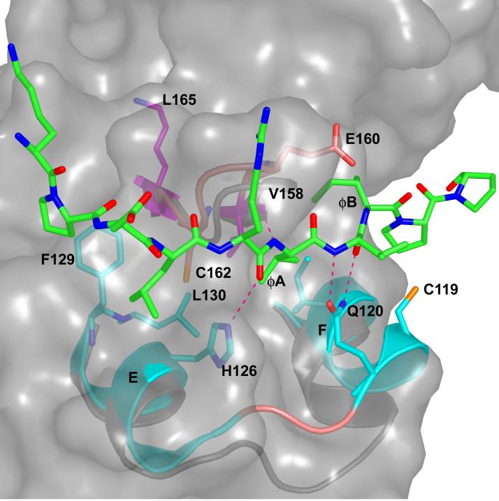
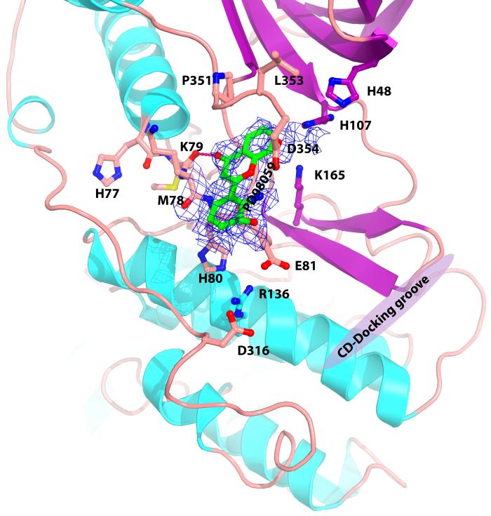
Map kinases are drug targets for autoimmune disease, cancer, and apoptosis-related diseases. Drug discovery efforts have developed MAP kinase inhibitors directed toward the ATP binding site and neighboring "DFG-out" site, both of which are targets for inhibitors of other protein kinases. On the other hand, MAP kinases have unique substrate and small molecule binding sites that could serve as inhibition sites. The substrate and processing enzyme D-motif binding site is present in all MAP kinases, and has many features of a good small molecule binding site. Further, the MAP kinase p38alpha has a binding site near its C-terminus discovered in crystallographic studies. Finally, the MAP kinases ERK2 and p38alpha have a second substrate binding site, the FXFP binding site that is exposed in active ERK2 and the D-motif peptide induced conformation of MAP kinases. Crystallographic evidence of these latter two binding sites is presented.


Figures

Fig. 1
Fig. 1
The structure of inactive p38α with substrate and small molecule binding sites indicated. The ATP binding site, the DFG-out site, CD-docking groove, FXFP binding site, and backside site are indicated as violet ellipsoids. Helices are cyan, β-strands magenta, and loops deep salmon. Figures generated using PyMOL (Delano Scientific, San Carlos, CA).
Fig. 2
Fig. 2
Docking groove interactions between p38α and pepMEF2A induced conformational changes. Cartoon of p38α docking groove; residues 110-129 + 157-163. Ø-2 through ØB of pepMEF2A is shown in green and all atom colors. The p38α/pepMEF2A is shown in cyan and all atom colors, and uncomplexed p38α is shown in gray. The side chains of I116, C119, Q120, H126, F129, L130, V158, and C162 and backbone of E160 p38α/pepMEF2A are displayed in ball and stick rendering. Dotted lines denote hydrogen bonds (2.6-3.2 Å). The surface of unliganded p38α in the docking groove is shown to reveal the full size of the pocket in the absence of peptide.
Fig. 3
Fig. 3
ERK2/pepHePTP reveals electron density near the FXFP binding site and peptide induced conformational changes. Electron density (|2Fo-Fc|, contoured at 0.9 σ) near the helix G and the MAP kinase insertion. Although the D-motif peptide (not shown) binds in the same docking groove as MEF2A binds to p38α [45], extra electron density is observed near helix G in ERK2. Ribbon diagram of the ERK2/pepHePTP (displayed between 176-203 +225-263 is in cyan. The side chains of L198, Y231, L232, and Y261 are shown in ball and stick. Uncomplexed ERK2 is shown in gray, and F181 and L182 are shown in ball and stick. Note that this binding pocket is available in ERK2/pepHePTP but not in ERK2.
Fig. 4
Fig. 4
Electron density of map with coefficients (|2Fo-Fc|) contoured at 0.7 σ for p38α crystals soaked in A) sulindac sulfide, and B) PD98059. The sulfur atom of sulindac sulfide is yellow, fluorine orange, oxygen red, and nitrogen blue. Residues forming contacts, K79, E81, H107, K165, P351 and L353 are displayed in ball and stick. Residues R136 and D316 in the CD domain are also displayed. Location of D-motif docking groove shown in magenta.
Fig. 4
Fig. 4
Electron density of map with coefficients (|2Fo-Fc|) contoured at 0.7 σ for p38α crystals soaked in A) sulindac sulfide, and B) PD98059. The sulfur atom of sulindac sulfide is yellow, fluorine orange, oxygen red, and nitrogen blue. Residues forming contacts, K79, E81, H107, K165, P351 and L353 are displayed in ball and stick. Residues R136 and D316 in the CD domain are also displayed. Location of D-motif docking groove shown in magenta.

References

    1. Chen Z, Gibson TB, Robinson F, Silvestro L, Pearson G, Xu B, Wright A, Vanderbilt C, Cobb MH. MAP kinases. Chem. Rev. 2001;101:2449–2476. - PubMed
    1. Johnson GL, Lapadat R. Mitogen-activated protein kinase pathways mediated by ERK, JNK, and p38 protein kinases. Science. 2002;298:1911–1912. - PubMed
    1. Deng Q, Liao R, Wu BL, Sun P. High intensity ras signaling induces premature senescence by activating p38 pathway in primary human fibroblasts. J. Biol. Chem. 2004;279:1050–1059. - PubMed
    1. Ferrell JE, Jr., Bhatt RR. Mechanistic studies of the dual phosphorylation of mitogen-activated protein kinase. J. Biol. Chem. 1997;272:19008–19016. - PubMed
    1. Pearson G, Robinson F, Beers Gibson T, Xu BE, Karandikar M, Berman K, Cobb MH. Mitogen-activated protein (MAP) kinase pathways: regulation and physiological functions. Endocr. Rev. 2001;22:153–183. - PubMed

Publication types

MeSH terms

Substances