. 2008 Jan 25;375(4):969-78.
doi: 10.1016/j.jmb.2007.11.013. Epub 2007 Nov 13.

Structure of antibody F425-B4e8 in complex with a V3 peptide reveals a new binding mode for HIV-1 neutralization

Affiliations

Structure of antibody F425-B4e8 in complex with a V3 peptide reveals a new binding mode for HIV-1 neutralization

Christian H Bell et al. J Mol Biol. .

Abstract

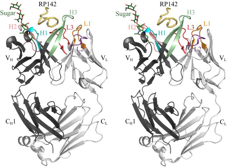
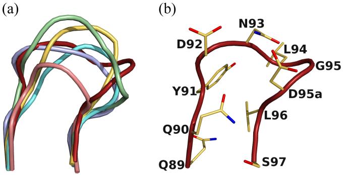
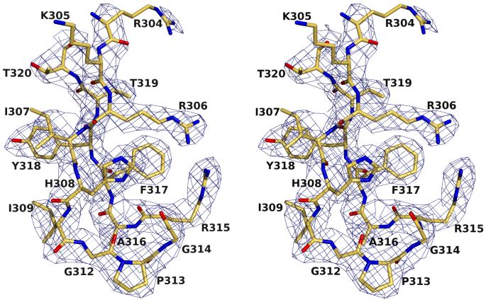
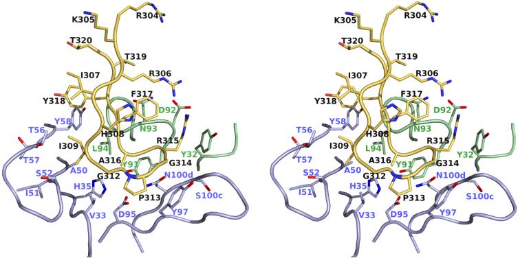
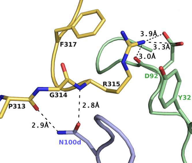
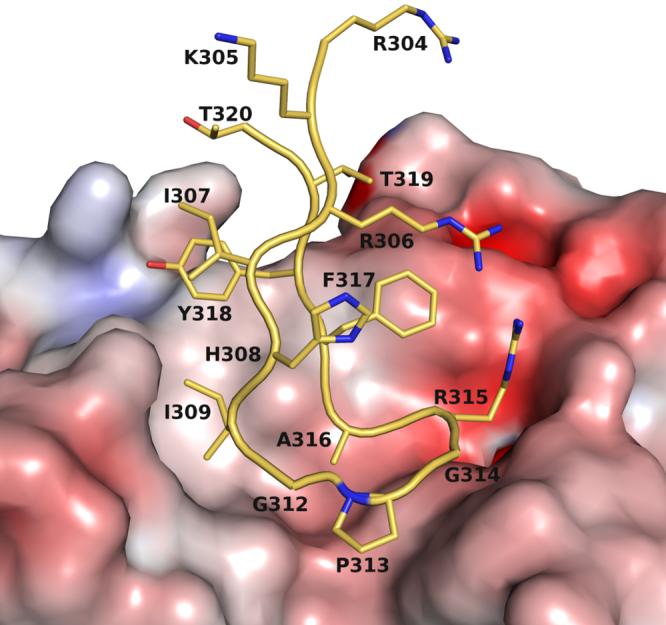
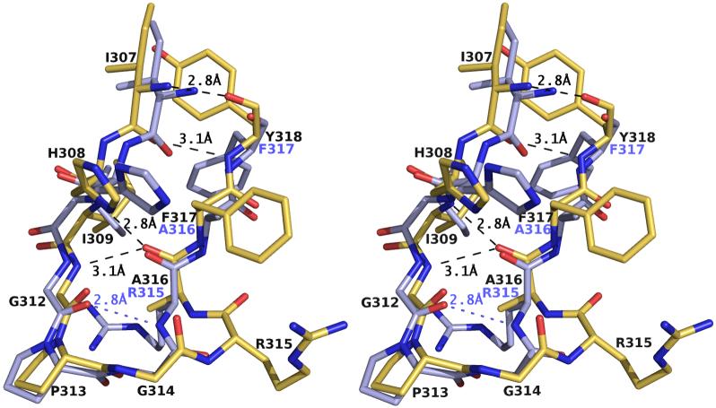
F425-B4e8 (B4e8) is a monoclonal antibody isolated from a human immunodeficiency virus type 1 (HIV-1)-infected individual that recognizes the V3 variable loop on the gp120 subunit of the viral envelope spike. B4e8 neutralizes a subset of HIV-1 primary isolates from subtypes B, C and D, which places this antibody among the very few human anti-V3 antibodies with notable cross-neutralizing activity. Here, the crystal structure of the B4e8 Fab' fragment in complex with a 24-mer V3 peptide (RP142) at 2.8 A resolution is described. The complex structure reveals that the antibody recognizes a novel V3 loop conformation, featuring a five-residue alpha-turn around the conserved GPGRA apex of the beta-hairpin loop. In agreement with previous mutagenesis analyses, the Fab' interacts primarily with V3 through side-chain contacts with just two residues, Ile(P309) and Arg(P315), while the remaining contacts are to the main chain. The structure helps explain how B4e8 can tolerate a certain degree of sequence variation within V3 and, hence, is able to neutralize an appreciable number of different HIV-1 isolates.


Figures

Figure 1
Figure 1
Crystal structure of the B4e8-RP142 complex. The peptide is colored in yellow and the Fab’ heavy and light chains in dark and light gray, respectively; the Fab’ CDR loops are colored in orange for L1, purple for L2, red for L3, cyan for H1, salmon for H2 and light green for H3. The sugar and AsnH28, to which it is linked, are shown in stick representation and colored in dark green. Images were produced with Pymol (http://pymol.sourceforge.net/).
Figure 2
Figure 2
New canonical class for CDR L3. (a) Comparison of the CDR L3 loop of B4e8 (red) to members of canonical class κ-1 (PDB-code: 1REI, lightblue), κ-2 (2FBJ, yellow), κ-3 (1YQV, aquamarine), κ-4 (1DFB, salmon) and κ-5 (palegreen, 1BAF). (b) The B4e8 kappa chain CDR L3 has an Asp instead of the usual Pro at position 95a and represents a new canonical structure.
Figure 3
Figure 3
Electron density (2Fobs-Fcalc) for the RP142-peptide in complex with B4e8, contoured at 1.0σ. All residues, except the side chains of LysP305 and ArgP304, are well defined.
Figure 4
Figure 4
Specific binding interactions between B4e8 and RP142. Atomic coloring is used for the atoms with the carbon atoms of the peptide colored in yellow, carbon atoms of the heavy chain in light blue and those of the light chain in pale green. Only those residues of the Fab’ that make contact to the peptide are shown in stick representation. Residues GlyP312 - AlaP316 form a five residue α-turn.
Figure 5
Figure 5
Detailed view of the exposed ArgP315 and its interactions with B4e8. Hydrogen bonds and salt bridges are shown by dashed lines. Color coding is as in Fig. 4. ArgP315 is sandwiched between PheP317 and TyrL32 and stabilized by a salt bridge and a hydrogen bond to the side chain and backbone of AspL92. The unusual α-turn conformation is stabilized by two hydrogen bonds between AsnH100d and the backbone of ArgP315 and ProP313, respectively.
Figure 6
Figure 6
Molecular surface representation of the antigen-binding pocket colored by electrostatic potential, calculated in APBS, and mapped onto the surface with the color code ranging from -10 kT/e (bright red) to +10 kT/e (dark blue). IleP309 is accommodated in a hydrophobic pocket, whereas ArgP315 forms a salt bridge with AspL92.
Figure 7
Figure 7
Comparison of V3-peptides RP142 and MN bound to B4e8 (atomic coloring with carbon atoms in yellow) and 2219 (carbon atoms in light blue), respectively. Identical residues in both peptides are labeled only for B4e8 (black). Identical hydrogen bonds are marked by dashed lines and shown only for B4e8. Due to the conversion from a type II β-turn in 2219 to an α-turn in B4e8, one hydrogen bond in the apex of RP142 is lost in the complex with B4e8. The β-hairpin conformation is maintained by a one-residue shift accompanied by a 180° rotation of AlaP316 and all subsequent residues. Residues P304 to P306 and P319 to 320 are omitted for clarity.

References

    1. Wyatt R, Sodroski J. The HIV-1 envelope glycoproteins: fusogens, antigens, and immunogens. Science. 1998;280:1884–1888. - PubMed
    1. Cardoso RM, Zwick MB, Stanfield RL, Kunert R, Binley JM, Katinger H, Burton DR, Wilson IA. Broadly neutralizing anti-HIV antibody 4E10 recognizes a helical conformation of a highly conserved fusion-associated motif in gp41. Immunity. 2005;22:163–173. - PubMed
    1. Zwick MB, Jensen R, Church S, Wang M, Stiegler G, Kunert R, Katinger H, Burton DR. Anti-human immunodeficiency virus type 1 (HIV-1) antibodies 2F5 and 4E10 require surprisingly few crucial residues in the membrane-proximal external region of glycoprotein gp41 to neutralize HIV-1. J. Virol. 2005;79:1252–1261. - PMC - PubMed
    1. Ofek G, Tang M, Sambor A, Katinger H, Mascola JR, Wyatt R, Kwong PD. Structure and mechanistic analysis of the anti-human immunodeficiency virus type 1 antibody 2F5 in complex with its gp41 epitope. J. Virol. 2004;78:10724–10737. - PMC - PubMed
    1. Saphire EO, Parren PW, Pantophlet R, Zwick MB, Morris GM, Rudd PM, Dwek RA, Stanfield RL, Burton DR, Wilson IA. Crystal structure of a neutralizing human IgG against HIV-1: a template for vaccine design. Science. 2001;293:1155–1159. - PubMed

Publication types

MeSH terms

Associated data