. 2008 Jun;12(3):229-39.
doi: 10.1016/j.media.2007.10.008. Epub 2007 Nov 6.

Preventing facial recognition when rendering MR images of the head in three dimensions

Affiliations

Preventing facial recognition when rendering MR images of the head in three dimensions

François Budin et al. Med Image Anal. 2008 Jun.

Abstract

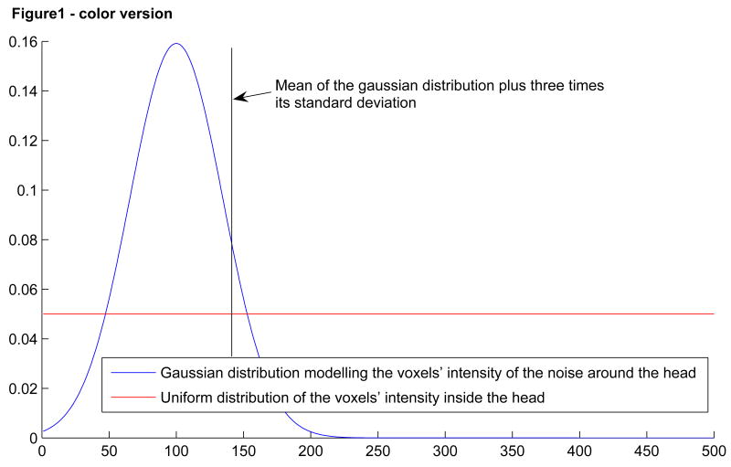
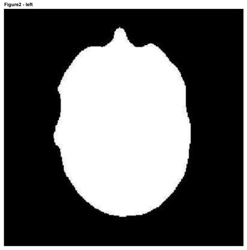
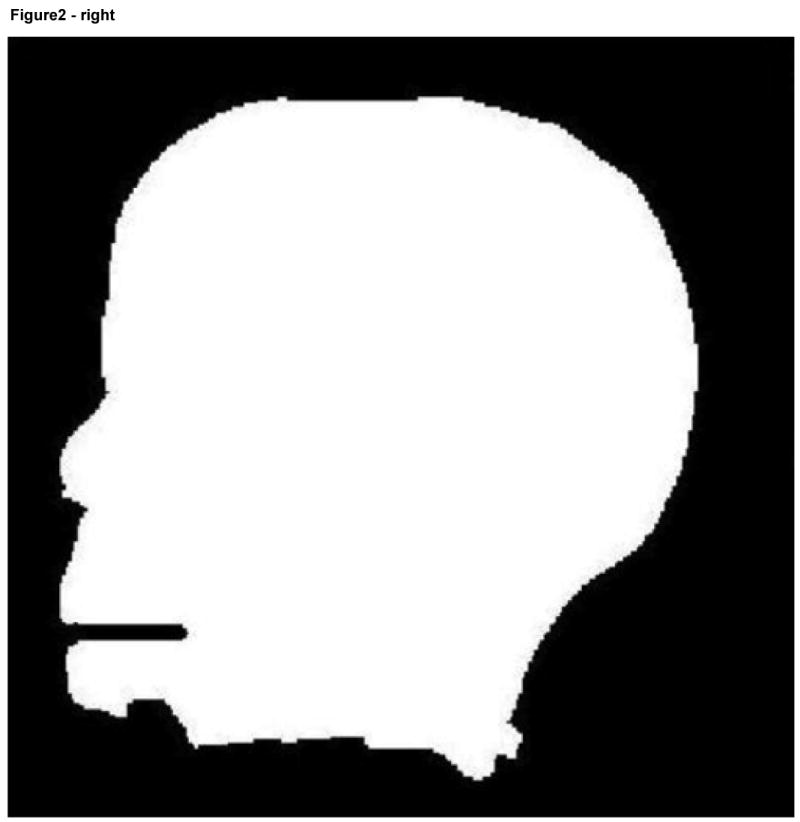
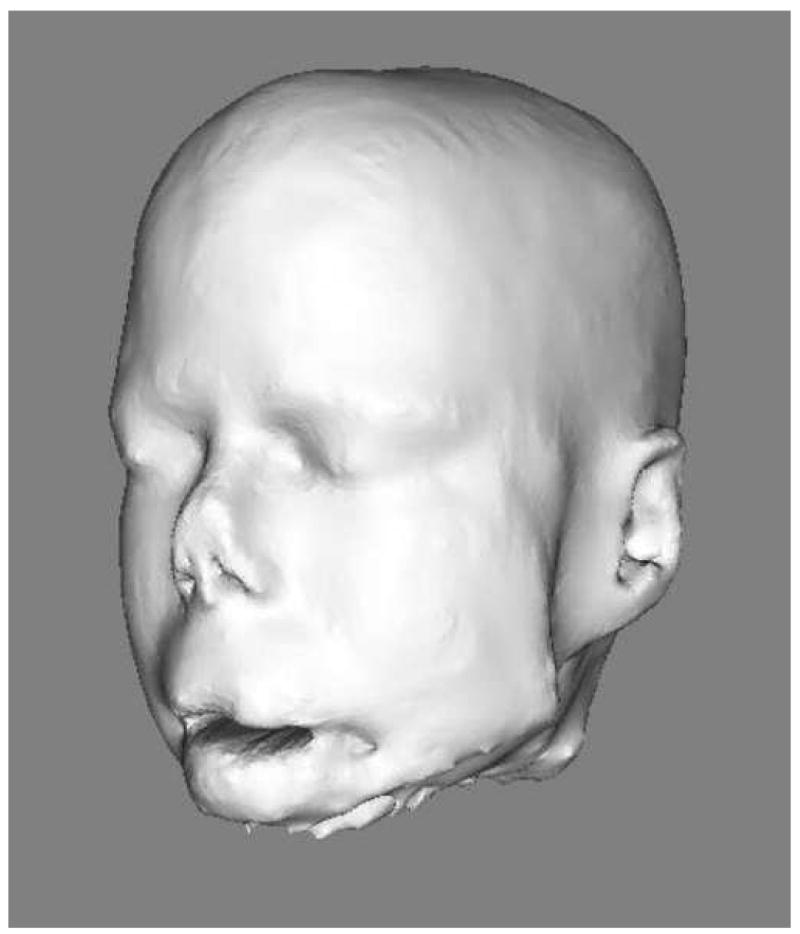
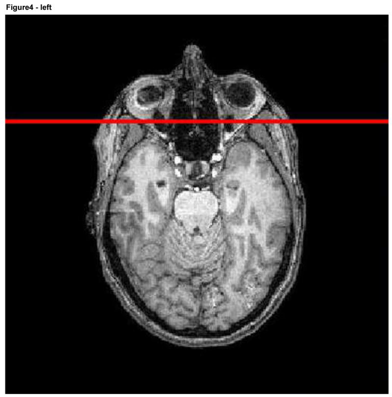
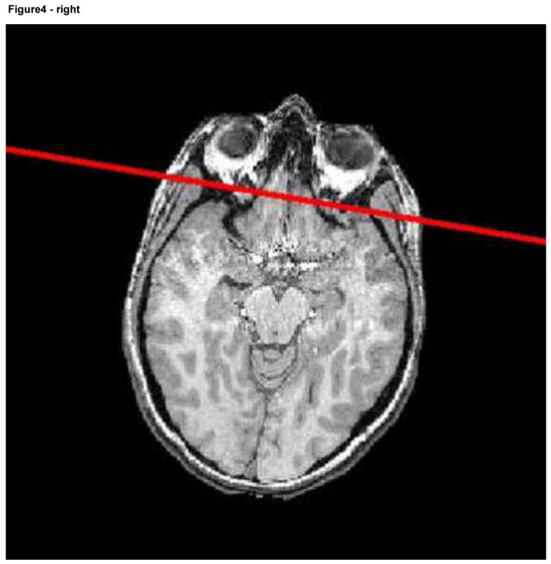
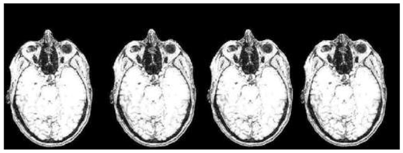
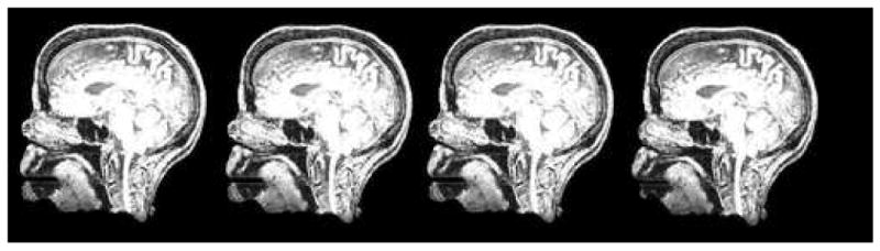
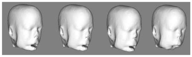
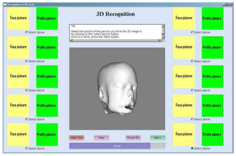
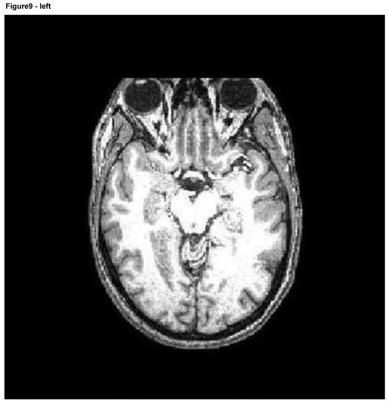
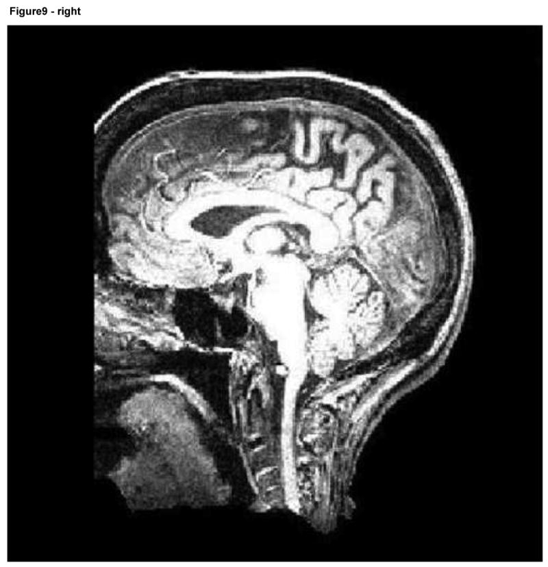
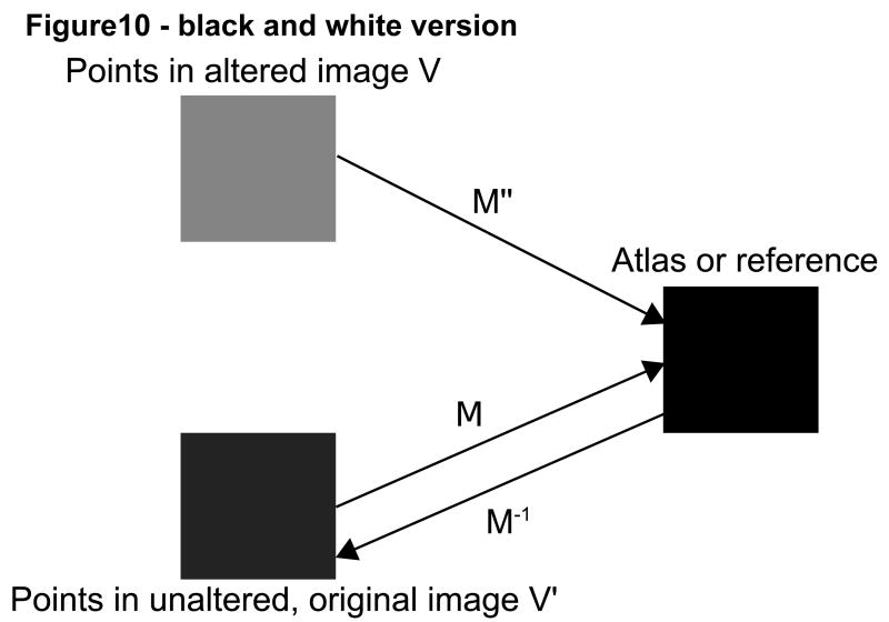
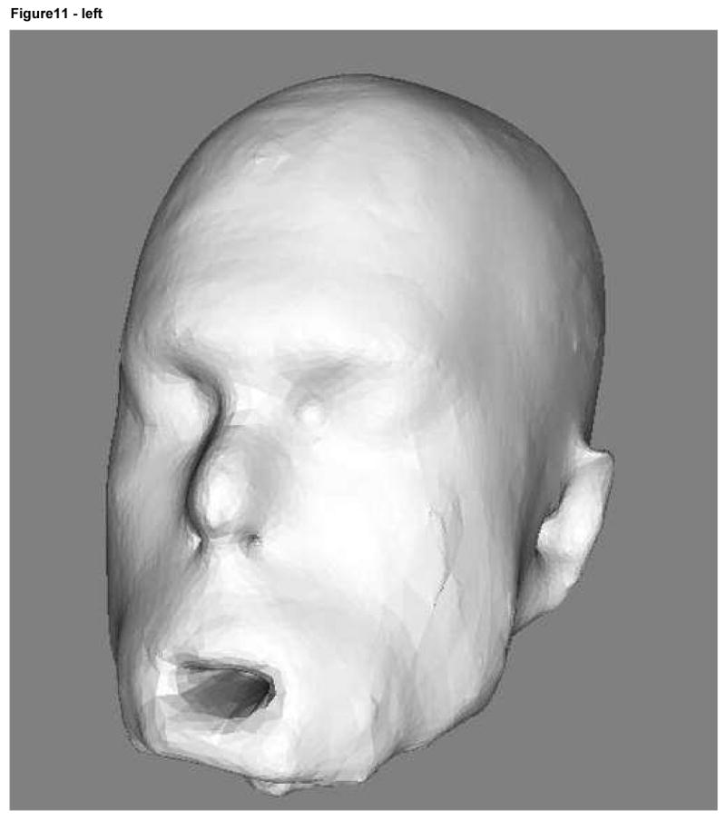
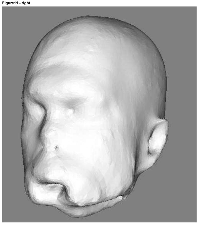
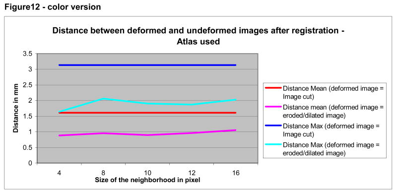
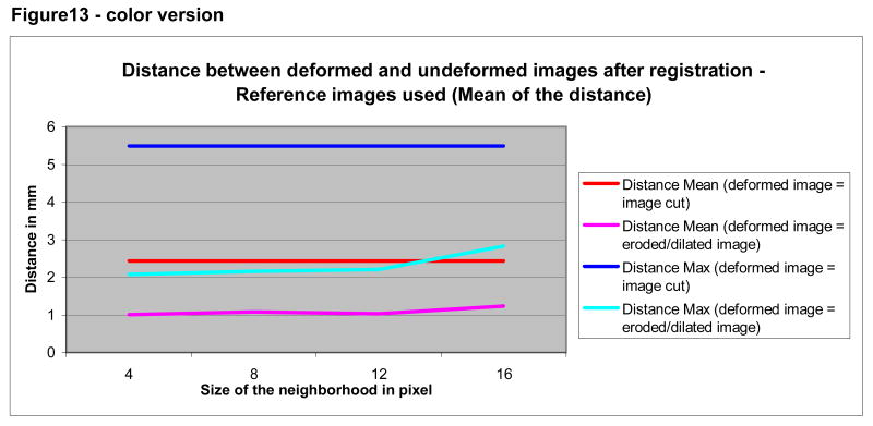
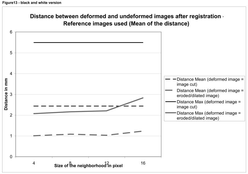
In the United States it is not allowed to make public any patient-specific information without the patient's consent. This ruling has led to difficulty for those interested in sharing three-dimensional (3D) images of the head and brain since a patient's face might be recognized from a 3D rendering of the skin surface. Approaches employed to date have included brain stripping and total removal of the face anterior to a cut plane, each of which lose potentially important anatomical information about the skull surface, air sinuses, and orbits. This paper describes a new approach that involves (a) definition of a plane anterior to which the face lies, and (b) an adjustable level of deformation of the skin surface anterior to that plane. On the basis of a user performance study using forced choices, we conclude that approximately 30% of individuals are at risk of recognition from 3D renderings of unaltered images and that truncation of the face below the level of the nose does not preclude facial recognition. Removal of the face anterior to a cut plane may interfere with accurate registration and may delete important anatomical information. Our new method alters little of the underlying anatomy and does not prevent effective registration into a common coordinate system. Although the methods presented here were not fully effective (one subject was consistently recognized under the forced choice study design even at the maximum deformation level employed) this paper may point a way toward solution of a difficult problem that has received little attention in the literature.


Figures

Figure 1
Figure 1
Model of the voxels' intensity distribution and the threshold extracted
Figure 1
Figure 1
Model of the voxels' intensity distribution and the threshold extracted
Figure 2
Figure 2
Axial (left) and sagittal (right) views of a binary facial mask.
Figure 2
Figure 2
Axial (left) and sagittal (right) views of a binary facial mask.
Figure 3
Figure 3
3D rendering of the head
Figure 4
Figure 4
Reference image with the associated plane shown in red (left) and a second image including the same plane following registration.
Figure 4
Figure 4
Reference image with the associated plane shown in red (left) and a second image including the same plane following registration.
Figure 5
Figure 5
Axial slice of a progressively deformed image with deformation produced by different kernel sizes (from left to right: 0, 4, 8, 12 voxels). Note the progressive shortening of the nose. The brain, skull, orbital structures, deep nasal structures, and pharynx remain unaltered.
Figure 6
Figure 6
Sagittal slice of the same set of images shown in Figure 5.
Figure 7
Figure 7
3D rendering of the same set of images shown in Figures 5 and 6.
Figure 8
Figure 8
View of the program used in the observer recognition study. Photographs are not shown because we do not have permission from all participants to use their photographs in a publication.
Figure 9
Figure 9
Axial and sagittal slices of an image following facial removal. Note the loss of information about the air sinuses, orbits, and anterior skull.
Figure 9
Figure 9
Axial and sagittal slices of an image following facial removal. Note the loss of information about the air sinuses, orbits, and anterior skull.
Figure 10
Figure 10
Graphical depiction of affine mappings between the original (V') and altered (V) images via registration to a common coordinate system (atlas or reference).
Figure 10
Figure 10
Graphical depiction of affine mappings between the original (V') and altered (V) images via registration to a common coordinate system (atlas or reference).
Figure 11
Figure 11
Subject 9 3D rendering at deformation level 2 (radius of 8 voxels) on the left and level 3 (radius of 12 voxels) on the right.
Figure 11
Figure 11
Subject 9 3D rendering at deformation level 2 (radius of 8 voxels) on the left and level 3 (radius of 12 voxels) on the right.
Figure 12
Figure 12
Mean and maximum distance between deformed and undeformed images according to method of deformation (erosion/dilation or removal of the face). Registration was to the ICBM atlas.
Figure 12
Figure 12
Mean and maximum distance between deformed and undeformed images according to method of deformation (erosion/dilation or removal of the face). Registration was to the ICBM atlas.
Figure 13
Figure 13
Mean and maximum distance between deformed and undeformed images according to method of deformation (erosion/dilation or removal of the face). Registration was to reference images of two different individuals.
Figure 13
Figure 13
Mean and maximum distance between deformed and undeformed images according to method of deformation (erosion/dilation or removal of the face). Registration was to reference images of two different individuals.

References

    1. Chellappa R, Wilson CL, Sirohey S. Human and Machine Recognition of Faces: A Survey. Proceeding of the IEEE. 1995;83:705–740.
    1. HIPAA Privacy Rules. 2006. Available at http://privacyruleandresearch.nih.gov/pr_08.asp.
    1. Ibañez L, Schroeder W, Ng L, Cates J. The ITK Software Guide. Second. Kitware Inc; 2005.
    1. ICBM Atlas, McConnell Brain Imaging Centre, Montréal Neurological Institute, McGill University, Montréal, Canada.

    1. Jomier J, Guyon JP, Aylward S, et al. The Spatial Object Viewers Toolkit. http://public.kitware.com/SOViewer.

Publication types