. 2008 Jan;237(1):209-15.
doi: 10.1002/dvdy.21399.

The macroPARP genes Parp-9 and Parp-14 are developmentally and differentially regulated in mouse tissues

Affiliations

The macroPARP genes Parp-9 and Parp-14 are developmentally and differentially regulated in mouse tissues

Antoinette Hakmé et al. Dev Dyn. 2008 Jan.

Abstract

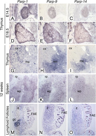
The macroPARPs Parp-9 and Parp-14 are macro domain containing poly(ADP-ribose) polymerases involved in transcriptional regulation in response to immunoregulatory cytokines. Their genes reside in the same locus (16B3), and the Parp-9 gene lies head-to-head and shares its promoter with the gene encoding its partner, Bbap. Here, we provide a detailed analysis of Parp-9, Parp-14, and Bbap expression during mouse development and adulthood. Parp-9 is developmentally regulated, and prominently expressed in the thymus and specific regions of the brain and gut. In adults, highest expression is maintained in the thymus and intestine. Parp-14 is more weakly expressed, mainly in the thymus during development and in adulthood. In addition, we show that Bbap is essentially coexpressed with Parp-9 during development and in adult mouse. However, the different levels of their transcripts detected in the developing brain and gut suggest that Bbap and Parp-9 display both common and independent tissue-specific regulations.


Figures

Figure 1
Figure 1
Expression of Parp‐1, Parp‐9, and Parp‐14 during mouse development. A–H: Sagittal sections of mouse embryos at embryonic day (E) 10.5 (A–C), 14.5 (D–F), and 17.5 (G,H) hybridized with antisense probes for Parp‐1 (A,D,G), Parp‐9 (B,E,H), Parp‐14 (F) or a control sense probe for Parp‐9 (C). bl, bladder; cc, cerebral cortex; cb, cerebellum; cl, cloaca; mc, mouth cavity; nc, nasal cavity; ob, olfactory bulb; pi, pituitary; pv, prevertebrae; re, rectum; te, telencephalic vesicle; th, thymus; to, tongue; us, urogenital sinus. The asterisk indicates a nonspecific signal (similarly observed with the sense probe, not shown).
Figure 2
Figure 2
Expression of Parp‐1, Parp‐9, and Parp‐14 in lymphoid organs during development and in adult mice. A–F: Sagittal sections of mouse embryos at embryonic day 14.5 (A–C), 18.5 (D–F) hybridized with antisense probes for Parp‐1 (A,D), Parp‐9 (B,E) and Parp‐14 (C,F). G–O: Sections of 12‐week‐old mouse thymus (G–I), spleen (J–L), and lymphoid follicles of distal colon (M–O) hybridized with antisense probes for Parp‐1 (G,J,M), Parp‐9 (H,K,N), Parp‐14 (I,L,O). cx, cortex; FAE, follicle associated epithelium; gc, germinal center; lf, lymphoid follicle; me, medulla; rp, red pulp; wp, white pulp.
Figure 3
Figure 3
Expression of Parp‐1, Parp‐9, and Parp‐14 in developing and adult mouse brain. A–I: Sagittal sections of mouse embryos at embryonic day 12.5 (A–C) and coronal sections of 12‐week‐old mouse brain hybridized with antisense probes for Parp‐1 (A,D,G), Parp‐9 (B,E,H), and Parp‐14 (C,F,I). CA1–3, hippocampal subregions; cc, cerebral cortex; dg, dentate gyrus; hb, hindbrain; nc, nasal cavity; te, telencephalic vesicle; pl, Purkinje cell layer.
Figure 4
Figure 4
Expression of Parp‐1, Parp‐9, and Parp‐14 in the developing and adult mouse intestine. A–I: Sagittal sections of mouse embryos at embryonic day 12.5 (A–C) or 18.5 (D,E), and sections of 12‐week‐old mouse ileum (F,G) and colon (H,I) hybridized with antisense probes for Parp‐1 (A,D,F,H), Parp‐9 (B,E,G,I), and Parp‐14 (C). co, colon; il, ileum; lu, lumen; ml, midgut loop; us, urogenital sinus.
Figure 5
Figure 5
Expression of Bbap during development and in adult mice. A–H: Sagittal sections of mouse embryos at embryonic day 10.5 (A) and 14.5 (B), and sections of 12‐week‐old mouse thymus (C), spleen (D), ileum (E), colon (F), and brain (G,H) hybridized with antisense probes for Bbap. CA1–3, hippocampal subregions; cc, cerebral cortex; cl, cloaca; cx, cortex; dg, dentate gyrus; hb, hindbrain; in, intestine; me, medulla; nc, nasal cavity; re, rectum; rp, red pulp; th, thymus te, telencephalic vesicle; pl, Purkinje cell layer; wp, white pulp.

Similar articles

Cited by

References

    1. Aguiar RC, Yakushijin Y, Kharbanda S, Salgia R, Fletcher JA, Shipp MA. 2000. BAL is a novel risk‐related gene in diffuse large B‐cell lymphomas that enhances cellular migration. Blood 96: 4328–4334. - PubMed
    1. Aguiar RC, Takeyama K, He C, Kreinbrink K, Shipp M. 2005. B‐aggressive lymphoma (BAL) family proteins have unique domains that modulate transcription and exhibit Poly(ADP‐ribose) polymerase activity. J Biol Chem 280: 33756–33765. - PubMed
    1. Amé JC, Spenlehauer C, de Murcia G. 2004. The PARP superfamily. Bioessays 26: 882–893. - PubMed
    1. Angelov D, Molla A, Perche PY, Hans F, Cote J, Khochbin S, Bouvet P, Dimitrov S. 2003. The histone variant macroH2A interferes with transcription factor binding and SWI/SNF nucleosome remodeling. Mol Cell 11: 1033–1041. - PubMed
    1. Artavanis‐Tsakonas S, Rand MD, Lake RJ. 1999. Notch signaling: cell fate control and signal integration in development. Science 284: 770–776. - PubMed

Publication types