. 2007 Dec;3(12):e174.
doi: 10.1371/journal.ppat.0030174.

A novel inhibitory mechanism of mitochondrion-dependent apoptosis by a herpesviral protein

Affiliations

A novel inhibitory mechanism of mitochondrion-dependent apoptosis by a herpesviral protein

Pinghui Feng et al. PLoS Pathog. 2007 Dec.

Abstract

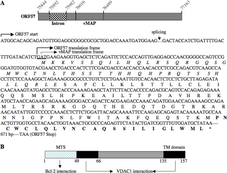
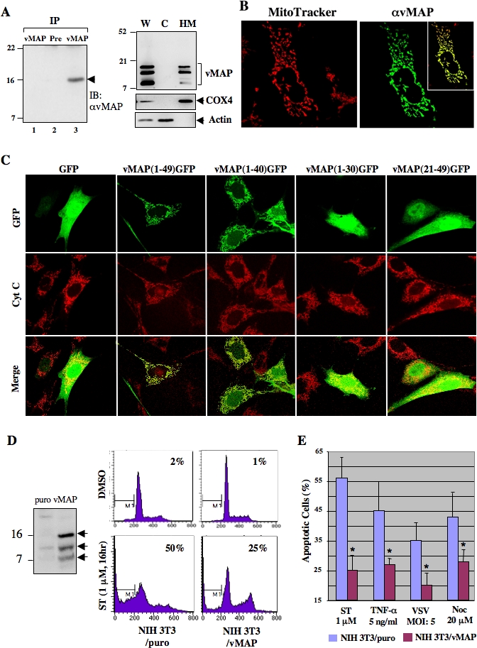
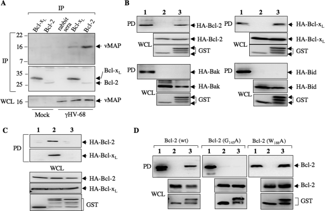
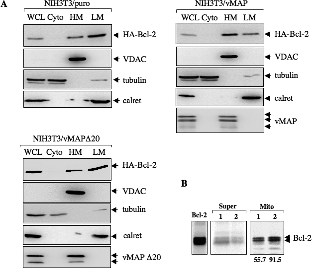
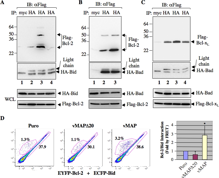
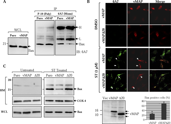
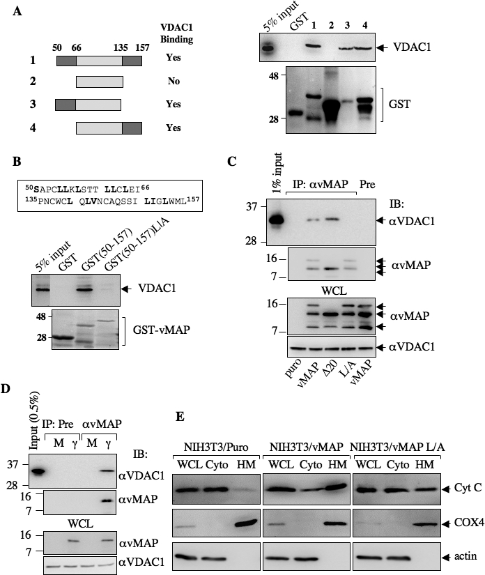
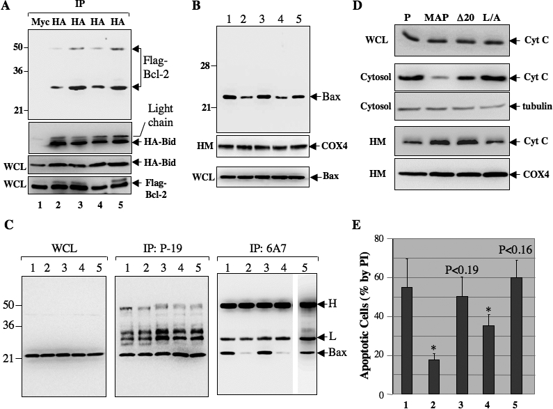
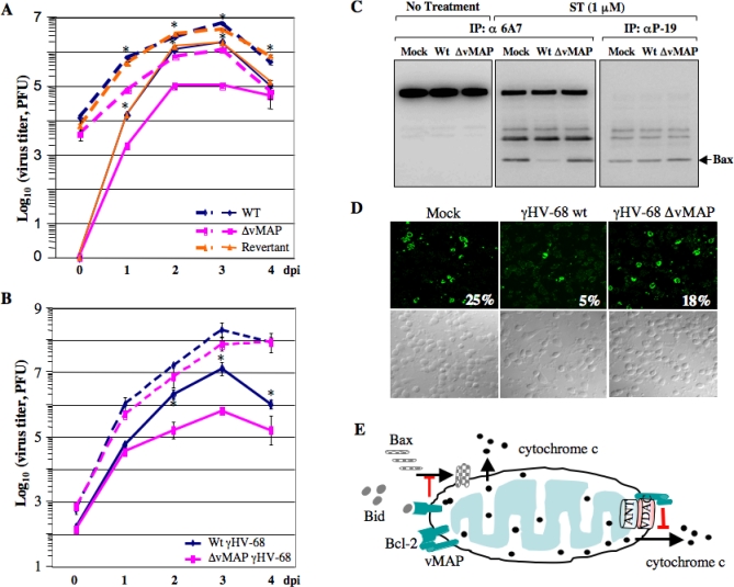
Upon viral infection, cells undergo apoptosis as a defense against viral replication. Viruses, in turn, have evolved elaborate mechanisms to subvert apoptotic processes. Here, we report that a novel viral mitochondrial anti-apoptotic protein (vMAP) of murine gamma-herpesvirus 68 (gammaHV-68) interacts with Bcl-2 and voltage-dependent anion channel 1 (VDAC1) in a genetically separable manner. The N-terminal region of vMAP interacted with Bcl-2, and this interaction markedly increased not only Bcl-2 recruitment to mitochondria but also its avidity for BH3-only pro-apoptotic proteins, thereby suppressing Bax mitochondrial translocation and activation. In addition, the central and C-terminal hydrophobic regions of vMAP interacted with VDAC1. Consequently, these interactions resulted in the effective inhibition of cytochrome c release, leading to the comprehensive inhibition of mitochondrion-mediated apoptosis. Finally, vMAP gene was required for efficient gammaHV-68 lytic replication in normal cells, but not in mitochondrial apoptosis-deficient cells. These results demonstrate that gammaHV-68 vMAP independently targets two important regulators of mitochondrial apoptosis-mediated intracellular innate immunity, allowing efficient viral lytic replication.


Conflict of interest statement

Competing interests. The authors have declared that no competing interests exist.

Figures

Figure 1
Figure 1. Genomic Localization and Protein Organization of γHV-68 vMAP
(A) vMAP is encoded within the ORF57. (Top diagram) The vMAP genomic localization is shown within the ORF57 sequence. Numbers indicate the nucleotide position according to Entrez accession number U97553. The grid box represents a 90–base pair intron of ORF57 and the box with vertical lines represents vMAP (M8). (Bottom diagram, sequence) The first ATG denotes the start codon for ORF57 (arrow on the left). The coding sequence of vMAP is shown in reference to ORF57 and its amino acid sequence is listed below. The translation of vMAP has a +1 shift to ORF57 frame; thus, the vMAP amino acid sequence is completely different from the ORF57 amino acid sequence. The mitochondrion-targeting sequence (MTS) is italicized and the putative transmembrane (TM) domain is indicated by bold letters. An arrow (↓) over a gap (dots) indicates the splicing site where a 90–base pair of intron is removed [47]. *, stop codon. (B) vMAP protein organization. The N terminal sequence (aa 1–49) serves as a mitochondrial targeting sequence (MTS) as well as a Bcl-2-interacting region. Both the internal hydrophobic region (aa 50–66) and the putative transmembrane (TM) domain (aa 135–157) interact with VDAC1.
Figure 2
Figure 2. γHV-68 vMAP Encodes a Mitochondrial Protein
(A) Identification of vMAP protein. (Left panel) γHV-68-infected (lane 2 and 3) or mock-infected NIH3T3 cells (lane 1) were harvested at 16 h post-infection. Post-centrifuged lysates in CHAPS buffer were precipitated with pre-immune serum (lane 2) or anti-vMAP serum (lanes 1 and 3), followed by immunoblotting (IB) with anti-vMAP serum. The arrowhead indicates vMAP. (Right panel) Subcellular fractionation of vMAP in 293T cells at 36 h post-transfection, as shown by immunoblotting with antibodies to vMAP, COX4, or actin. W, whole cell lysate; C, cytosolic; HM, mitochondrion-enriched heavy membrane. (B) vMAP localizes to the mitochondrion. Post-transfection with pcDNA5-vMAP vector, COS-1 cells were stained with MitoTracker and fixed for anti-vMAP immunostaining. The box inside of the right panel shows the merged image of MitoTracker and anti-vMAP immunostaining. (C) Identification of the vMAP MTS. Either GFP alone or various vMAP N-terminal sequences fused to GFP were expressed in NIH3T3 cells. Mitochondria were stained with anti–cytochrome c antibody (red). Pictures represent more than 85% of cells analyzed by fluorescent microscopy. (D) vMAP inhibits apoptosis. NIH3T3/puro and NIH3T3/vMAP cells were treated with DMSO or ST (1 μM) for 16 h and stained with PI, followed by flow cytometry analysis. Data are from one of three replicate experiments. vMAP (indicated by arrows) expression was shown by immunoblotting on the left. (E) vMAP inhibits apoptosis initiated by various apoptogenic stimuli. NIH3T3/puro and NIH3T3/vMAP cells were treated with various agents (ST, TNF-α/cycloheximide, vesicular stomatitis [VSV] infection for 16 h, or nocodazole [Noc] for 36 h), and sub-G1 cells were quantified as described in (D). Data represent result of three independent experiments and error bars indicate standard deviation with (*) p < 0.05 relative to control (puro) as calculated by Student's t-test.
Figure 3
Figure 3. vMAP Interacts with Bcl-2/Bcl-xL
(A) vMAP interacts with Bcl-2/Bcl-xL in γHV-68-infected cells. At 16 h post-infection, γHV-68-infected (MOI = 1) or mock-infected NIH3T3 cells were harvested and post-centrifuge supernatants were subjected to immunoprecipitation (IP) with rabbit antibodies to Bcl-2 or Bcl-xL, followed by immunoblotting with anti-vMAP serum. A normal rabbit serum was included as a negative control. Protein precipitates were analyzed by immunoblotting with antibodies to Bcl-2 and Bcl-xL (middle panel). Whole cell lysates (WCL) of mock-infected or γHV-68-infected NIH3T3 cells were immunoblotted with anti-vMAP serum (bottom panel). (B) The N-terminal sequence of vMAP interacts with Bcl-2/Bcl-xL but not with Bak and Bid. At 48 h post-transfection with a plasmid expressing HA-Bcl-2, HA-Bcl-xL, HA-Bak, or HA-Bid, and a plasmid expressing GST (lane 2) or vMAP(1–50)-GST (lane 3), cell lysates of 293T were used for GST pull-down (PD) assay, followed by immunoblotting with anti-HA (top panel of each set). WCLs were analyzed by immunoblotting with antibodies to HA epitope (Bcl-2 family proteins, middle panels) and GST (bottom panels) for GST or vMAP(1–50)-GST expression. Lane 1 is 2% input of lysates of cells expressing GST and Bcl-2 family proteins. For all four sets of PDs (top panel), lane 1 shows the equivalent amount of Bcl-2 family proteins compared to those shown in the middle panel. (C) The first 20 amino acids of vMAP are essential for its interaction with Bcl-2/Bcl-xL. 293T cells were transfected with a plasmid expressing HA-Bcl-2, HA-Bcl-xL together with a plasmid expressing GST (lane 1), vMAP-GST (lane 2), or vMAPΔ20-GST (lane 3). Mammalian GST pull-down was carried out as described in (B). The precipitates were analyzed by immunoblotting with anti-HA to detect Bcl-2 and Bcl-xL (top two panels). WCLs were analyzed by immunoblotting with anti-HA and anti-GST antibodies to demonstrate the expression of Bcl-2/Bcl-xL and GST fusion, respectively. (D) The Bcl-2 BH2 domain is not involved in vMAP binding. At 48 h post-transfection with a plasmid expressing HA-Bcl-2, Bcl-2 G145A, or Bcl-2 W188A together with a plasmid expressing GST (lane 2) or vMAP(1–50)-GST (lane 3), 293T cell lysates were used for GST pull-down, followed by immunoblotting with Bcl-2 antibody (top panel of each set). WCLs were analyzed by immunoblotting with antibodies to Bcl-2 (middle panel) and GST (bottom panel). Lane 1 indicates 2% input of lysates of cells expressing vMAP-GST and Bcl-2/Bcl-xL.
Figure 4
Figure 4. vMAP Enhances the Mitochondrial Localization of Bcl-2
(A) Bcl-2 intracellular distribution by subcellular fractionation. NIH3T3/puro, NIH3T3/vMAP, and NIH3T3/vMAPΔ20 cells were transfected with HA-Bcl-2 expression vector. Twenty μg of WCL, cytosolic (Cyto), heavy membrane (HM), and light membrane (LM) fractions were analyzed by immunoblotting with antibodies to HA epitope (HA-Bcl2, top panel), VDAC, tubulin, calreticulin (calret), or vMAP. (B) In vitro Bcl-2 mitochondrial association assay. The mitochondrion-enriched HM fractions of NIH3T3/puro (lane 1) or NIH3T3/vMAP cells (lane 2) were mixed with in vitro translated 35S-labeled Bcl-2 at 30 °C for 2 h, centrifuged at 13,000 rpm for 15 min to separate soluble (Super) and insoluble (Mito), followed by autoradiograph. The left panel indicates Bcl-2 translated in rabbit reticulocyte lysates. The numbers below autography indicate the levels (intensity of the Bcl-2 doublet) of mitochondrion-associated Bcl-2 determined by Phosphor Imager analysis.
Figure 5
Figure 5. vMAP Enhances the Interaction between Bcl-2/Bcl-xL and BH3-Only Proteins
(A–C) NIH3T3/puro cells (lane 2), NIH3T3/vMAP cells (lanes 1 and 3), and NIH3T3/vMAPΔ20 cells (lane 4) were transfected with plasmids expressing HA-Bid and Flag-Bcl-2 (A), HA-Bad and Flag-Bcl-2 (B), or HA-Bad and Flag-Bcl-xL (C). WCLs were used for immunoprecipitation with a mouse monoclonal anti-HA antibody, followed by immunoblotting with HRP-conjugated anti-Flag antibody to detect Flag-Bcl-2 and Flag-Bcl-xL (top panel) or with anti-HA antibody to detect the precipitated HA-Bid and HA-Bad (second panels). WCLs were used to detect HA-Bid, HA-Bad, Flag-Bcl-2, and Flag-Bcl-xL expression (bottom two panels) using antibodies to the HA epitope or Flag epitope. Lane 1 indicates anti-Myc immunoprecipitation of lysates from NIH3T3/vMAP cells as a negative control. The light chains of the Myc and HA antibodies migrate differently on SDS-PAGE. (D) Fluorescence resonance energy transfer (FRET) assay. At 36 h post-transfection with pEYFP-Bcl-2 and pECFP-Bid, NIH3T3/puro cells (Puro, panel 1), NIH3T3/vMAPΔ20 cells (vMAPΔ20, panel 2), and NIH3T3/vMAP cells (vMAP, panel 3) were harvested and subjected to flow cytometry analysis. Numbers in the FRET diagrams indicate the percentage of the transfected cell population without protein interaction (lower right) and the transfected cell population with protein interaction (upper left). The right panel shows the data in the graph representing an average from two independent experiments, and error bars indicate standard deviation with (*) p < 0.05 relative to control (Puro) as calculated by Student's t-test.
Figure 6
Figure 6. vMAP Inhibits the Mitochondrial Translocation and Activation of Bax
(A) vMAP inhibits the apoptotic conformational change of Bax. NIH3T3/puro (Puro) and NIH3T3/vMAP (vMAP) cells were treated with ST (1 μM) for 4 h, harvested, and lysed in 1% CHAPS buffer. Pre-cleared cell lysates were split into two fractions, and each was used for immunoprecipitation with the mouse 6A7 monoclonal antibody or the rabbit P-19 polyclonal antibody, followed by immunoblotting with 6A7. WCLs were analyzed by immunoblotting with 6A7 antibody. Of note, 6A7 antibody reacts only with the activated Bax in immunoprecipitation and immunofluorescence assays, but with pan-bax in the immunoblotting assay. H and L indicate the heavy and light chains of immunoglobulin, respectively. WCL, whole cell lysate. (B) vMAP inhibits Bax activation by immunofluorescence assay. At 16 h post-transfection with a plasmid expressing vMAP or vMAPΔ20, HeLa cells were treated with DMSO or ST (1 μM) for 4 h, fixed, and stained with rabbit anti-vMAP serum (red) and the mouse 6A7 monoclonal anti-Bax antibody (green). A single representative optical section is presented. Arrows indicate cells expressing vMAP or vMAPΔ20. The bottom right graph represents data collected from over 200 transfected cells. Error bars indicate standard deviation with (*) p = 0.01 relative to vMAPΔ20 as calculated by Student's t-test. The bottom left panel shows the expression of vMAP and vMAPΔ20 (Δ20). (C) vMAP inhibits Bax translocation by fractionation. NIH3T3/puro cells (Puro), NIH3T3/vMAP cells (vMAP), or NIH3T3/vMAPΔ20 cells (Δ20) were untreated or treated with ST (1 μM) for 4 h, and the mitochondrion-enriched HM fractions (20 μg) were resolved by SDS-PAGE and analyzed by immunoblotting with antibodies to detect endogenous Bax and COX4. WCLs were analyzed by immunoblotting with anti-Bax antibody (bottom panel).
Figure 7
Figure 7. vMAP Interaction with VDAC1 Inhibits Cytochrome c Release
(A) vMAP contains two VDAC1-interacting regions (dark grey boxes). The left panel shows the schematic diagram of vMAP interaction with VDAC1. (Right panel) GST fusions containing various vMAP sequences were expressed and purified from E. coli. 293T cells were lysed in CHAPS buffer and subjected to in vitro GST pull-down, followed by immunoblotting with anti-VDAC antibody. The bottom panel shows the Coomassie blue staining of GST fusion proteins. Lanes 1–4 correspond to the GST fusions shown by the left diagram. (B) The LLxL repeat sequences of vMAP are required for VDAC1 interaction. (Top box) The boxed sequences are the two LLxL repeats of vMAP. GST or GST fusion proteins were used to bind to VDAC1 from 293T cell lysates in CHAPS buffer as described in (A). Protein precipitates were analyzed by immunoblotting with anti-VDAC antibody (top blot) and Coomassie blue staining of GST fusion proteins (bottom blot). (C) vMAP interacts with VDAC1 in stable cell lines. NIH3T3 cells stably expressing vector, vMAP, or its mutants were used for immunoprecipitation with anti-vMAP serum or pre-immune (Pre) serum, followed by immunoblotting with anti-VDAC antibody (top panel) or anti-vMAP serum (middle panel). WCLs were analyzed by immunoblotting with anti-vMAP serum and anti-VDAC antibody (bottom two panels). (D) vMAP interacts with VDAC1 in γHV-68-infected cells. WCLs of mock (M)- or γHV-68 (γ)-infected (MOI = 1) cells were precipitated with anti-vMAP or preimmune serum, followed by immunoblotting with anti-VDAC antibody (top panel) or anti-vMAP serum (middle panel). WCLs were analyzed by immunoblotting with anti-vMAP serum and anti-VDAC antibody (bottom two panels). (E) vMAP inhibits cytochrome c release upon ST treatment. NIH3T3/puro, NIH3T3/vMAP, or NIH3T3/vMAP L/A cells were treated with ST (1 μM) for 4 h and WCLs were sequentially centrifuged to obtain cytosolic (Cyto) and mitochondrion-enriched heavy membrane (HM) fractions. Polypeptides (20 μg) from each fraction were analyzed by immunoblotting with antibodies to cytochrome c (Cyt C), COX4, and actin.
Figure 8
Figure 8. vMAP Interacts with Bcl-2 and VDAC1 in a Genetically Separable Manner
(A) vMAP interaction with Bcl-2, but not with VDAC1, enhances Bcl-2 binding to Bid. NIH3T3 stable cells expressing puro (lane 2), vMAP (lanes 1 and 3), vMAPΔ20 (lane 4), or vMAP L/A (lane 5) were transfected with plasmids containing Flag-Bcl-2 and HA-Bid. At 48 h post-transfection, cells were harvested and WCLs were used for immunoprecipitation with anti-HA (Bid), followed by immunoblotting with HRP-conjugated anti-Flag (Bcl-2, top panel) or anti-HA antibody (middle panel). WCLs were analyzed by immunoblotting with anti-HA (Bid) and anti-Flag (Bcl-2) antibodies (bottom two panels). Anti-Myc antibody in lane 1 was included as a negative control. (B) vMAP-Bcl-2 interaction but not vMAP-VDAC1 interaction is required to inhibit Bax mitochondrial translocation. NIH3T3 stable cells were treated with ST (1 μM) for 4 h, and mitochondrion-enriched HMs (20 μg) were analyzed by immunoblotting with anti-Bax or anti-VDAC antibody (top two panels). WCLs were used for immunoblotting with anti-Bax (bottom panel). Lane 1, NIH3T3/puro; lane 2, NIH3T3/vMAP; lane 3, NIH3T3/vMAPΔ20; lane 4, NIH3T3/vMAP L/A; lane 5, NIH3T3/vMAPΔ20&L/A. (C) vMAP-Bcl-2 interaction but not vMAP-VDAC1 interaction is required to inhibit the mitochondrial translocation of Bax. NIH3T3 cells expressing wt vMAP or mutants thereof were stimulated with ST (1 μM) for 4 h. WCLs were precipitated with anti-Bax P-19 polyclonal antibody or 6A7 monoclonal antibody, followed by immunoblotting with 6A7 monoclonal antibody as described in Figure 6A. The content of the lanes is same as described in (B). H and L indicate the heavy and light chains of immunoglobulin, respectively. (D) vMAP interactions with Bcl-2 and VDAC1 are required to efficiently inhibit cytochrome c release. NIH3T3 cells containing vector (P), vMAP, vMAPΔ20 (Δ20), or vMAP L/A (L/A) were treated with ST (1 μM) for 4 h. Mitochondrion-enriched HM and cytosolic fractions (Cytosol) were obtained by centrifugation as described in Materials and Methods. Each fraction (20 μg) was resolved by SDS-PAGE and analyzed by immunoblotting with antibodies to cytochrome c (Cyt C), COX4, and tubulin (cytosolic fraction only). (E) Roles of vMAP interactions with Bcl-2 and VDAC1 in anti-apoptosis. NIH3T3 cells were treated with ST (1 μM) for 16 h and subsequently stained with PI for flow cytometry. Data represent three independent experiments. Error bars indicate standard deviation with (*) p < 0.05 relative to control (NIH3T3/puro cells, 1) as calculated by Student's t-test. The content of the lanes is same as described in (B).
Figure 9
Figure 9. vMAP Anti-Apoptotic Activity Is Required for the Efficient Replication of γHV-68
(A) vMAP is required for the efficient replication of γHV-68 in NIH3T3 cells. NIH3T3 cells were infected with γHV-68 wt, γHV-68 ΔvMAP, or γHV-68 Rev at MOI = 0.01 (solid line) or MOI = 5 (dashed line). Culture (cells and supernatants) was harvested at various time points post-infection. Plaque assay was performed with BHK21 cells. Data represent duplicate experiments and error bars indicate standard deviation with (*) p < 0.07 relative to γHV-68 ΔvMAP as calculated by Student's t-test. dpi, days post infection. The p-value can be applied for both γHV-68 wt and γHV-68 Rev. (B) vMAP is dispensable for γHV-68 replication in Bax−/−Bak−/− (DKO) MEF. wt or Bax−/−Bak−/− (DKO) MEF cells were infected with γHV-68 wt or γHV-68 ΔvMAP at MOI = 1. Culture (cells and supernatants) was harvested at various time points post infection. Solid lines and dashed lines indicate growth kinetics of γHV-68 in wt MEFs and DKO MEFs, respectively. Data represent the results of two independent measurements and error bars indicate standard deviation with (*) p < 0.06 relative to wt MEF as calculated by Student's t-test. (C and D) vMAP expression inhibits Bax activation during γHV-68 replication. NIH3T12 cells were mock-infected (Mock), infected with γHV-68 wt (Wt) or γHV-68 ΔvMAP (ΔvMAP) at MOI = 2 for 12 h, and then untreated or treated with ST at 1 μM for 4 h. Bax activation was examined by immunoprecipitation (P-19 and 6A7) and immunoblotting (6A7) (C) or confocal microscopy with 6A7 monoclonal antibody (D) as described in Figure 5. Numbers in (D) indicate the percentages of 6A7 Bax conformer positive cells. (E) A hypothetical model of vMAP action in the inhibition of mitochondrion-mediated apoptosis. γHV-68 vMAP recruits Bcl-2 to the mitochondrion and enhances Bcl-2 interaction with BH3-only proteins, thereby blocking Bax translocation and activation. On the other hand, vMAP interactions with both Bcl-2 and VDAC1 lead to a comprehensive inhibition of cytochrome c release upon apoptotic stress. Red-colored lines indicate vMAP-mediated inhibition.

Similar articles

Cited by

References

    1. Susin SA, Lorenzo HK, Zamzami N, Marzo I, Snow BE, et al. Molecular characterization of mitochondrial apoptosis-inducing factor. Nature. 1999;397:441–446. - PubMed
    1. Li P, Nijhawan D, Budihardjo I, Srinivasula SM, Ahmad M, et al. Cytochrome c and dATP-dependent formation of Apaf-1/caspase-9 complex initiates an apoptotic protease cascade. Cell. 1997;91:479–489. - PubMed
    1. Du C, Fang M, Li Y, Li L, Wang X. Smac, a mitochondrial protein that promotes cytochrome c-dependent caspase activation by eliminating IAP inhibition. Cell. 2000;102:33–42. - PubMed
    1. Verhagen AM, Ekert PG, Pakusch M, Silke J, Connolly LM, et al. Identification of DIABLO, a mammalian protein that promotes apoptosis by binding to and antagonizing IAP proteins. Cell. 2000;102:43–53. - PubMed
    1. Kuwana T, Mackey MR, Perkins G, Ellisman MH, Latterich M, et al. Bid, Bax, and lipids cooperate to form supramolecular openings in the outer mitochondrial membrane. Cell. 2002;111:331–342. - PubMed

Publication types

MeSH terms

Substances