. 2007 Dec;3(12):e218.
doi: 10.1371/journal.pgen.0030218.

A RNA interference screen identifies the protein phosphatase 2A subunit PR55gamma as a stress-sensitive inhibitor of c-SRC

Affiliations

A RNA interference screen identifies the protein phosphatase 2A subunit PR55gamma as a stress-sensitive inhibitor of c-SRC

Pieter J A Eichhorn et al. PLoS Genet. 2007 Dec.

Abstract

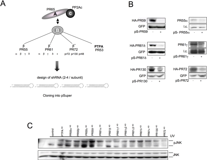
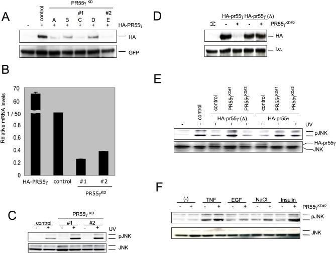
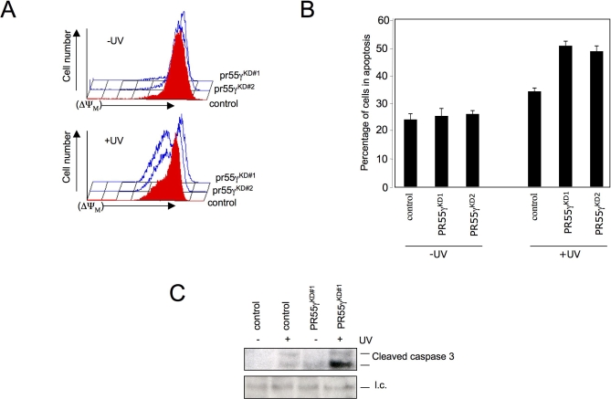
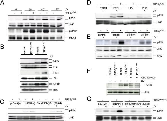
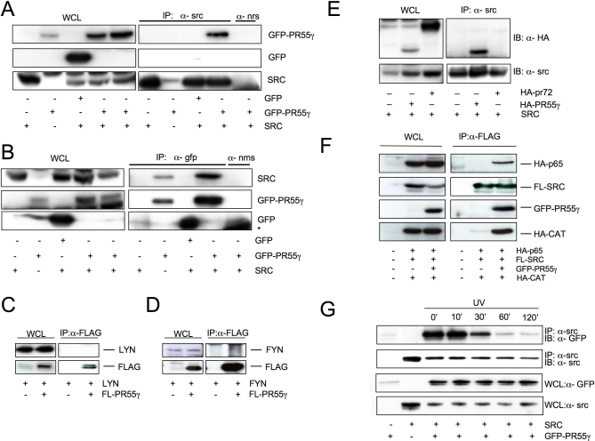
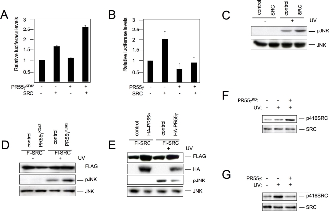
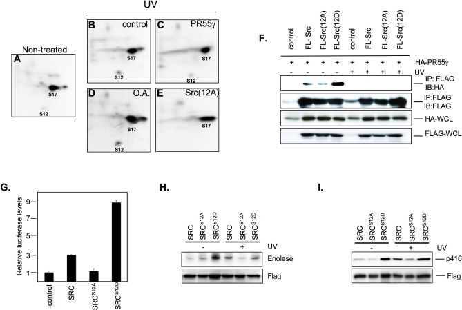
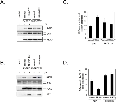
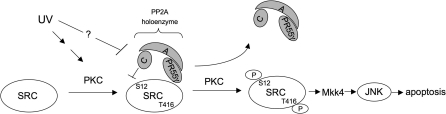
Protein Phosphatase type 2A (PP2A) represents a family of holoenzyme complexes with diverse biological activities. Specific holoenzyme complexes are thought to be deregulated during oncogenic transformation and oncogene-induced signaling. Since most studies on the role of this phosphatase family have relied on the use of generic PP2A inhibitors, the contribution of individual PP2A holoenzyme complexes in PP2A-controlled signaling pathways is largely unclear. To gain insight into this, we have constructed a set of shRNA vectors targeting the individual PP2A regulatory subunits for suppression by RNA interference. Here, we identify PR55gamma and PR55delta as inhibitors of c-Jun NH(2)-terminal kinase (JNK) activation by UV irradiation. We show that PR55gamma binds c-SRC and modulates the phosphorylation of serine 12 of c-SRC, a residue we demonstrate to be required for JNK activation by c-SRC. We also find that the physical interaction between PR55gamma and c-SRC is sensitive to UV irradiation. Our data reveal a novel mechanism of c-SRC regulation whereby in response to stress c-SRC activity is regulated, at least in part, through loss of the interaction with its inhibitor, PR55gamma.


Conflict of interest statement

Competing interests. The authors have declared that no competing interests exist.

Figures

Figure 1
Figure 1. PP2A Family Screen
(A) Schematic of the PP2A holoenzyme and outline of the B regulatory subunit families. (B) U2-OS cells were transfected with the indicated pSuper constructs and where available cotransfected with an HA-tagged version of the corresponding PP2A B subunit. Immunoblot panels show the efficiency of knockdown in six different pools as judged by the ability to knockdown cotransfected or endogenous protein (GFP is a transfection control). (C) U2-OS cells were cotransfected with pooled PP2A shRNAs or a control vector. Levels of phosphorylated JNK (α-pJNK) or JNK1 and JNK2 (α-JNK) were shown in cell lysates for the different samples 60 min after UV treatment of the cells. doi:10.1371/journal.pgen.0030218.g001
Figure 2
Figure 2. PR55γ Is a Regulator of JNK following UV Irradiation
(A) U2-OS cells expressing pcDNA-HA-PR55γ were cotransfected with the pooled knockdown (PR55γKD) vectors as indicated (A–E) or a control vector. GFP expression serves as a measure of transfection consistency. (B) U2-OS cells were cotransfected with PR55γKD vectors #1 or #2, pcDNA-PR55γ serves as a positive control. pSuper vector targeting a mouse PP2A subunit PR59 served as an shRNA control. mRNA levels relative to the control are shown as evaluated by quantitative real-time PCR. (C) U2-OS cells were cotransfected with PR55γKD vectors as indicated (#1 or #2) or control vector. Selected cells were exposed to UV irradiation (100 J/m2) and incubated for a further 60 min. Protein samples were analyzed by immunoblotting with antibodies targeting phosphorylated JNK (α-pJNK) or JNK1 and JNK2 (α-JNK). (D) U2-OS cells expressing pcDNA-HA-PR55γ or pcDNA-HA-PR55γ (Δ) were cotransfected with PR55γKD vector #2. Protein samples were analyzed by immunoblotting with antibodies targeting HA. (E) U2-OS cells expressing pcDNA-HA-PR55γ, pcDNA-HA- PR55γ(Δ), or a control vector were cotransfected with PR55γKD vectors #1 or #2. A pSuper vector targeting a mouse PP2A subunit PR59 served as an shRNA control. Selected cells were exposed to UV irradiation (100 J/m2) and incubated for a further 60 min. Protein samples were analyzed by immunoblotting with antibodies targeting phosphorylated JNK (α- pJNK), total JNK (α-JNK), or haemoglutinin (α-HA, reprobe). (F) U2-OS cells expressing PR55γKD2 vector or a control vector exposed to TNF-α, EGF, NaCl, or insulin for 5 min and incubated for a further 30–60 minutes. pJNK relative to total JNK levels are shown. doi:10.1371/journal.pgen.0030218.g002
Figure 3
Figure 3. PR55γ Is a Regulator of Apoptosis following UV Irradiation
(A) U2-OS cells expressing PR55γ shRNAs as indicated. Selected cells were exposed to UV irradiation (50 J/m2) and treated 18 h later with fluorescent dye measuring mitochondria membrane potential (DiOC6[3)]. FACS scan analysis of treated cells is shown. Right and left peaks reflect living and apoptotic cells, respectively. Top panel graph represents breakdown of the mitochondrial membrane potential in the absence of UV. Bottom panel graph represents breakdown of the mitochondrial membrane potential in the presence of UV. (B) Shown is the percentage of apoptotic cells treated with or without UV. Figure represents three independent experiments. (C) U2-OS cells were cotransfected with PR55γKD vector #2 or control vector. Selected cells were exposed to UV irradiation (50 J/m2) and incubated for a further 3 h. Protein samples were analyzed by immunoblotting with antibodies targeting phosphorylated cleaved caspase 3. A background band denotes loading control. doi:10.1371/journal.pgen.0030218.g003
Figure 4
Figure 4. PR55γ Regulates JNK Upstream of MKK4 and at the Level or Upstream of c-SRC
(A) pJNK and pMKK4 in relation to the total level of unphosphorylated protein in UV irradiated U2-OS cells followed over time (0–60 min) in the presence or absence of PR55γKD2 vector. (B) U2-OS cells expressing PR55γKD2 or a control vector were treated with UV and incubated for 60 min. Whole cell extracts were probed with the indicated antibodies. (C) PR55γKD2 vector or control vector were cotransfected as indicated in the presence or absence of c-SRC295M (dominant negative). Selected cells were exposed to UV irradiation and whole-cell extracts were analyzed by immunoblotting with antibodies targeting phosphorylated JNK (α-pJNK) or JNK1 and JNK2 (α-JNK). (D) U2-OS cells were cotransfected with PR55γKD2 vector or control vector as indicated and incubated with PP2 for 2 h and UV for 1 h. Whole cell extracts were analyzed by immunoblotting with antibodies targeting phosphorylated JNK (α- pJNK) or JNK1 and JNK2 (α-JNK). (E) PR55γKD2 vector or control vector were cotransfected as indicated in the presence or absence of pSuper-c-SRC. Selected cells were exposed to UV irradiation and whole cell extracts were analyzed by immunoblotting with antibodies targeting phosphorylated JNK (α- pJNK), JNK1 and JNK2 (α-JNK), or SRC(α- SRC). (F) PR55γKD2 vector or control vector were cotransfected as indicated in the presence or absence of CDC42V12 (dominant active). Selected cells were exposed to UV irradiation and whole cell extracts were analyzed by immunoblotting with antibodies targeting phosphorylated JNK (α-pJNK) or JNK1 (α-JNK). (G) Cells were transfected with either the PP2A pool targeting PR55δ vector or control vector and cotransfected as indicated in the presence or absence of c-SRC295M (dominant negative). Selected cells were exposed to UV irradiation and whole cell extracts were analyzed by immunoblotting with antibodies targeting phosphorylated JNK (α-pJNK) or JNK1 and JNK2 (α-JNK). doi:10.1371/journal.pgen.0030218.g004
Figure 5
Figure 5. SRC Coimmunoprecipitates with PR55γ
(A) U2-OS cells were transfected as indicated. At 48 h after transfection cells were lysed in ELB and c-SRC was immunoprecipitated using a polyclonal rabbit c-SRC antibody or normal rabbit serum (nrs). Coimmunoprecipitated GFP-tagged PR55γ was detected with a GFP antibody. Whole cell lysates (WCL) were probed using c-SRC or GFP antibodies to detect transfected protein. (B) Reverse immunoprecipitation as in (A) GFP was immunoprecipitated using a mouse GFP antibody or normal mouse serum (nms). Coimmunoprecipitated c-SRC was detected with a c-SRC specific antibody. The blot was subsequently reprobed with a GFP antibody to visualize the amount of immunoprecipitated GFP-tagged PR55γ protein. Whole cell lysates (WCL) were probed using c-SRC or GFP antibodies to detect transfected protein. (*) represents the light chain antibody. (C) U2-OS cells were transfected as indicated. Lysates were immunoprecipitated using a FLAG antibody. The blot was probed with a LYN specific antibody and then subsequently reprobed with a FLAG antibody to visualize the amount of immunoprecipitated FLAG-tagged PR55γ protein. (D) U2-OS cells were transfected as indicated. Lysates were immunoprecipitated using a FLAG antibody. The blot was probed with a FYN specific antibody and then subsequently reprobed with a FLAG antibody to visualize the amount of immunoprecipitated FLAG-tagged PR55γ protein. (E) U2-OS were transfected as indicated. c-SRC was immunoprecipitated using a c-SRC antibody. Coimmunoprecipitated HA-tagged PR55γ or HA-tagged PR72 was detected with a HA-specific antibody. The blot was subsequently reprobed with a c-SRC antibody to visualize the amount of immunoprecipitated c-SRC protein. Whole cell lysates (WCL) were probed using c-SRC or HA antibodies to detect transfected protein. (F) Immune precipitation of FLAG-tagged c-SRC and the PP2Ac/PR65 core dimer in HEK 293 cells in the presence (right) or absence (left) of GFP-PR55γ. The left panel shows 10% of the total lysate. (G) U2-OS cells were transfected as indicated. At 48 h after transfection cells were treated with UV (100 J/ m2) and incubated for a further 10, 30, 60, and 120 min, respectively. Equal amounts of cell lysate (500 μg) were immunoprecipitated with α- c-SRC antibody. Coimmunoprecipitated GFP-tagged PR55γ was detected with a GFP specific antibody. The blot was subsequently reprobed with a c-SRC antibody to visualize the amount of immunoprecipitated c-SRC protein. Whole cell lysates (WCL) were probed using c-SRC or GFP antibodies to detect transfected protein. doi:10.1371/journal.pgen.0030218.g005
Figure 6
Figure 6. PR55γ Regulates c-SRC-Induced JNK Activation
(A) U2-OS cells expressing a 5× AP1-luciferase construct (pGL2), pSUPER-PR55γ, or pcDNA-c-SRC as indicated. Luciferase counts are shown relative to the control. (B) U2-OS cells expressing a 5× AP1-luciferase construct (pGL2), pcDNA-HA-PR55γ, or pcDNA-c-SRC as indicated. Luciferase counts are shown relative to the control. (C) U2-OS cells were transfected with FLAG-SRC or a control vector and treated with UV. Whole cell extracts were probed with the indicated antibodies. (D) U2-OS cells were cotransfected with FLAG-SRC, pSUPER-PR55γ, or a control vector and treated with UV. Whole cell extracts were probed with the indicated antibodies. (E) U2-OS cells were cotransfected with FLAG-SRC, pcDNA-HA-PR55γ, or a empty vector and treated with UV. Whole cell extracts were probed with the indicated antibodies. (F) U2-OS cells expressing pcDNA-HA-hairpins targeting PR55γ or a control vector, and pcDNA-c-SRC were serum starved for 48 h and treated with UV irradiation for 30 min. Cells were lysed in ELB and equivalent amounts of protein were immunoprecipitated with a c-SRC specific antibody. c-SRC phosphorylation was detected with an antibody targeting phosphorylated tyrosine 416 (α-pTyr416) and immunoprecipitated c-SRC was detected with an α-c-SRC antibody. (G) U2-OS cells expressing pcDNA-HA-PR55γ or a control vector and pcDNA-c-SRC were serum starved for 48 h and treated with UV irradiation for 30 min. Cells were lysed in ELB and equivalent amounts of protein were immunoprecipitated with a c-SRC specific antibody. c-SRC phosphorylation was detected with an antibody targeting phosphorylated tyrosine 416 (α-pTyr416) and immunoprecipitated c-SRC was detected with an α-c-SRC antibody. doi:10.1371/journal.pgen.0030218.g006
Figure 7
Figure 7. PR55γ Regulates Serine 12 Phosphorylation on c-SRC
(A) Phosphotryptic pattern of U2-OS cells expressing c-SRC from untreated cells. (B–E) Phosphopeptides of c-SRC following treatment with UV (100 J/ m2) for 30 min in the presence of 2 mCi [32P]orthophosphate (B), c-SRC plus PR55γ (C), c-SRC pretreated for 1 h with 1 μM okadaic acid (D), and Ser12A mutant (E). Phosphorylated c-SRC was immunoprecipitated, and samples were subjected to phosphopeptide mapping as described under “Materials and Methods”. (F) U2-OS cells were transfected as indicated. 48 h after transfection cells were treated with UV (100 J/m2) and incubated for a further 120 min. Cells were lysed in ELB, and equal amounts of cell lysate (500 μg) were immunoprecipitated using a FLAG antibody (Sigma). Coimmunoprecipitated HA-tagged PR55γ was detected with an HA antibody. Whole cell lysates (WCL) were probed using FLAG or HA antibodies to detect transfected protein. (G) U2-OS cells expressing a 5× AP1-luciferase construct (pGL2), Flag-SRC, Flag- SRCS12A, Flag- SRCS12D, or control vector as indicated. Luciferase counts are shown relative to the control. (H) U2-OS cells were transfected as indicated. At 48 h after transfection cells were washed twice in PBS and incubated for a further 72 h in medium without FCS. Cells were treated with UV (200 J/m2) and incubated for a further 30 min in the same medium. Equal amounts of cell lysate (500 μg) were immunoprecipitated with α-Flag antibody and in vitro kinase assays were performed using enolase as a substrate. (I) U2-OS cells were transfected as indicated. At 48 h after transfection cells were washed twice in PBS and incubated for a further 72 h in medium without FCS. Cells were treated with UV (200 J/m2) and incubated for a further 30 min in the same medium. Equal amounts of cell lysate (500 μg) were immunoprecipitated with α-Flag antibody and analyzed by western blot for either tyrosine phosphorylation at residue 416 (top panel) or FLAG (bottom panel). doi:10.1371/journal.pgen.0030218.g007
Figure 8
Figure 8. Inhibition of JNK Activity by PR55γ Is Dependent upon Ser12 of c-SRC
(A) PR55γKD2 vector or control vector were cotransfected as indicated in the presence of FLAG-SRC or FLAG- SRCS12A. Selected cells were exposed to UV irradiation and whole cell extracts were analyzed by immunoblotting with antibodies targeting phosphorylated JNK (α-pJNK), JNK1 and JNK2 (α-JNK), or FLAG (α-FLAG). (B) GFP-PR55γ vector or control vector were cotransfected as indicated in the presence of FLAG-SRC or FLAG- SRCS12D. Selected cells were exposed to UV irradiation and whole cell extracts were analyzed by immunoblotting with antibodies targeting phosphorylated JNK (α- pJNK), JNK1 and JNK2 (α-JNK), FLAG (α-FLAG), or GFP (α-GFP). (C) U2-OS cells coexpressing PR55γ shRNAs and FLAG-SRC or FLAG- SRCS12A as indicated. Selected cells were exposed to UV irradiation (50 J/m2) and treated 18 h later with fluorescent dye measuring mitochondria membrane potential (DiOC6[3]). Figure represents three independent experiments. (D) U2-OS cells expressing PR55γ and FLAG-SRC or FLAG- SRCS12D as indicated. Selected cells were exposed to UV irradiation (50 J/m2) and treated 18 h later with fluorescent dye measuring mitochondria membrane potential (DiOC6[3]). Figure represents three independent experiments. doi:10.1371/journal.pgen.0030218.g008
Figure 9
Figure 9. Model for the Regulation of c-SRC-Induced JNK Activation following UV Signaling by PP2A Complexes
doi:10.1371/journal.pgen.0030218.g009

References

    1. Parsons SJ, Parsons JT. Src family kinases, key regulators of signal transduction. Oncogene. 2004;23:7906–7909. - PubMed
    1. Boggon TJ, Eck MJ. Structure and regulation of Src family kinases. Oncogene. 2004;23:7918–7927. - PubMed
    1. Zheng XM, Wang Y, Pallen CJ. Cell transformation and activation of pp60c-src by overexpression of a protein tyrosine phosphatase. Nature. 1992;359:336–339. - PubMed
    1. Bjorge JD, Pang A, Fujita DJ. Identification of protein-tyrosine phosphatase 1B as the major tyrosine phosphatase activity capable of dephosphorylating and activating c-Src in several human breast cancer cell lines. J Biol Chem. 2000;275:41439–41446. - PubMed
    1. Irby RB, Yeatman TJ. Role of Src expression and activation in human cancer. Oncogene. 2000;19:5636–5642. - PubMed

Publication types

MeSH terms