

Anatomic distribution of apoptosis in medulla oblongata of infants and adults
- PMID: 18069990
- PMCID: PMC2408973
- DOI: 10.1111/j.1469-7580.2007.00842.x
Anatomic distribution of apoptosis in medulla oblongata of infants and adults
Abstract
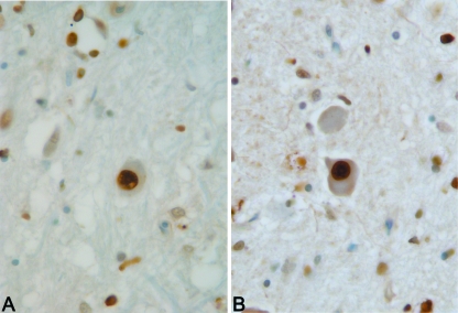
The aim of the study was to evaluate the distribution of apoptosis in the medullary nuclei of infants and adults who died of hypoxic-ischaemic injury. Apoptosis was studied by terminal deoxynucleotidyl transferase-mediated dUTP nick end-labelling (TUNEL) in brainstems from 22 adults (7 subjects who died of opiate intoxication, 15 who died of other hypoxic-ischaemic injury) and 10 infants. The nuclei examined included the hypoglossal, dorsal motor nucleus of the vagus, nucleus tractus solitarii, nucleus of the spinal trigeminal tract, cuneate, vestibular and inferior olivary nuclei. A morphometric analysis with the optical disector method was performed to calculate the mean percentages (+/- standard deviation) of TUNEL-positive neuronal and glial cells for the sample populations. Opiate deaths did not have higher apoptotic indices than other adult hypoxic-ischaemic deaths. Statistically significant differences between adults and infants were found in the neuronal apoptotic indices of the cuneate (28.2 +/- 16.3% vs. 6.9 +/- 8.7%), vestibular (24.7 +/- 15.0% vs. 11.3 +/- 11.4%), nucleus tractus solitarii (11.2 +/- 11.2% vs. 2.3 +/- 2.4%), dorsal motor nucleus of the vagus (6.8 +/- 8.5% vs. 0.1 +/- 0.2%) and hypoglossal (6.6 +/- 5.7% vs. 0.1 +/- 0.2%), indicating higher resistance of the neuronal populations of these infant medullary nuclei to terminal hypoxic-ischaemic injury or post-mortem changes. Differences in neuronal apoptotic index were also statistically significant among nuclei, suggesting differential characteristics of survival. Nuclei with higher neuronal apoptotic indices were the cuneate, vestibular and nucleus of the spinal trigeminal tract, which are located in the lateral medullary tegmentum and share the same vascular supply from the posterior inferior cerebellar artery.
Figures


Similar articles
-
Detection of apoptosis in human brainstem by TUNEL assay.Ital J Anat Embryol. 2005 Oct-Dec;110(4):255-60. Ital J Anat Embryol. 2005. PMID: 16536056
-
Morphometric analysis of infant and adult medullary nuclei through optical disector method.Anat Rec (Hoboken). 2009 Oct;292(10):1619-29. doi: 10.1002/ar.20957. Anat Rec (Hoboken). 2009. PMID: 19685502
-
Protective action of tetramethylpyrazine on the medulla oblongata in rats with chronic hypoxia.Auton Neurosci. 2013 Jan;173(1-2):45-52. doi: 10.1016/j.autneu.2012.11.004. Epub 2012 Dec 4. Auton Neurosci. 2013. PMID: 23218834
-
The dorsal motor nucleus of the vagus (DMNV) in sudden infant death syndrome (SIDS): pathways leading to apoptosis.Respir Physiol Neurobiol. 2013 Jan 15;185(2):203-10. doi: 10.1016/j.resp.2012.09.001. Epub 2012 Sep 10. Respir Physiol Neurobiol. 2013. PMID: 22975482 Review.
-
Serotonin- and substance P-containing projections to the nucleus tractus solitarii of the rat.J Comp Neurol. 1987 Nov 8;265(2):275-93. doi: 10.1002/cne.902650210. J Comp Neurol. 1987. PMID: 2447131 Review.
Cited by
-
Neuronal apoptosis in the brainstem medulla of sudden unexpected death in infancy (SUDI), and the importance of standardized SUDI classification.Forensic Sci Med Pathol. 2018 Mar;14(1):42-56. doi: 10.1007/s12024-018-9954-1. Epub 2018 Feb 19. Forensic Sci Med Pathol. 2018. PMID: 29460253
-
Pathogenesis of cognitive dysfunction in patients with obstructive sleep apnea: a hypothesis with emphasis on the nucleus tractus solitarius.Sleep Disord. 2012;2012:251096. doi: 10.1155/2012/251096. Epub 2012 Jan 16. Sleep Disord. 2012. PMID: 23470865 Free PMC article.
-
Cell death as a regulator of cerebellar histogenesis and compartmentation.Cerebellum. 2011 Sep;10(3):373-92. doi: 10.1007/s12311-010-0222-5. Cerebellum. 2011. PMID: 20941559 Review.
-
Death by a thousand cuts in Alzheimer's disease: hypoxia--the prodrome.Neurotox Res. 2013 Aug;24(2):216-43. doi: 10.1007/s12640-013-9379-2. Epub 2013 Feb 12. Neurotox Res. 2013. PMID: 23400634 Review.
-
Dysfunctional nucleus tractus solitarius: its crucial role in promoting neuropathogenetic cascade of Alzheimer's dementia--a novel hypothesis.Neurochem Res. 2012 Apr;37(4):846-68. doi: 10.1007/s11064-011-0680-2. Epub 2012 Jan 5. Neurochem Res. 2012. PMID: 22219130 Review.
References
-
- Adle-Biassette H, Levy Y, Colombel M, Poron F, Natchev S, Keohane C, et al. Neuronal apoptosis in HIV infection in adults. Neuropathol Appl Neurobiol. 1995;21:218–227. - PubMed
-
- Anglade P, Vyas S, Hirsch EC, Agid Y. Apoptosis in dopaminergic neurons of the human substantia nigra during normal aging. Histol Histopathol. 1997;12:603–610. - PubMed
-
- Atici S, Cinel L, Cinel I, Doruk N, Aktekin M, Akca A, et al. Opioid neurotoxicity: comparison of morphine and tramadol in an experimental rat model. Int J Neurosci. 2004;114:1001–1011. - PubMed
-
- Basheer R, Yang J, Tempel A. Chronic prenatal morphine treatment decreases G alpha s mRNA levels in neonatal frontal cortex. Brain Res Dev Brain Res. 1992;70:145–148. - PubMed
-
- Chan WY, Yew DT. Apoptosis and bcl-2 oncoprotein expression in the human fetal central nervous system. Anat Rec. 1998;252:165–175. - PubMed
MeSH terms
Substances
LinkOut - more resources
Full Text Sources
