Comparative Study
. 2008 Mar;49(3):400-9.
doi: 10.1111/j.1528-1167.2007.01458.x. Epub 2007 Dec 6.

Early treatment suppresses the development of spike-wave epilepsy in a rat model

Affiliations
Comparative Study

Early treatment suppresses the development of spike-wave epilepsy in a rat model

Hal Blumenfeld et al. Epilepsia. 2008 Mar.

Abstract

Purpose: Current treatments for epilepsy may control seizures, but have no known effects on the underlying disease. We sought to determine whether early treatment in a model of genetic epilepsy would reduce the severity of the epilepsy phenotype in adulthood.

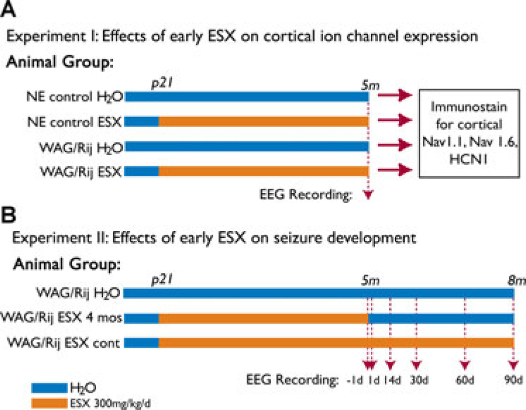
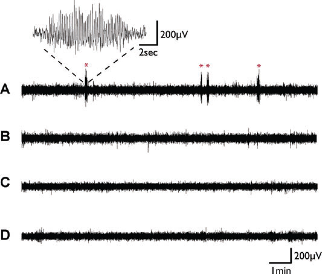
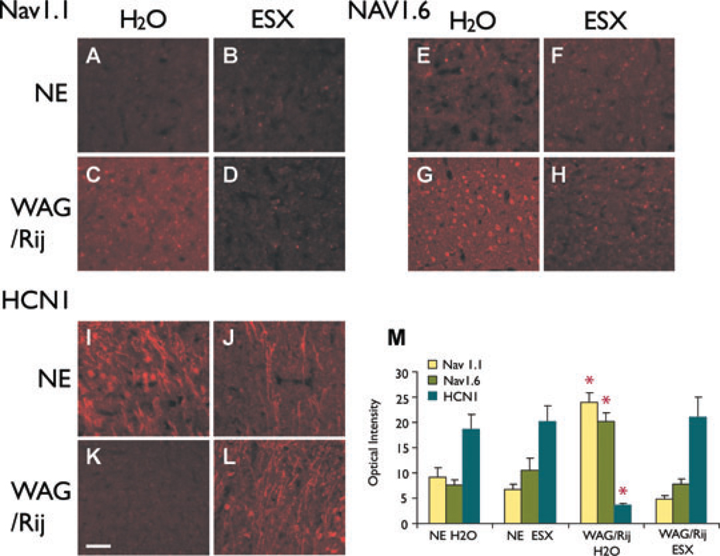
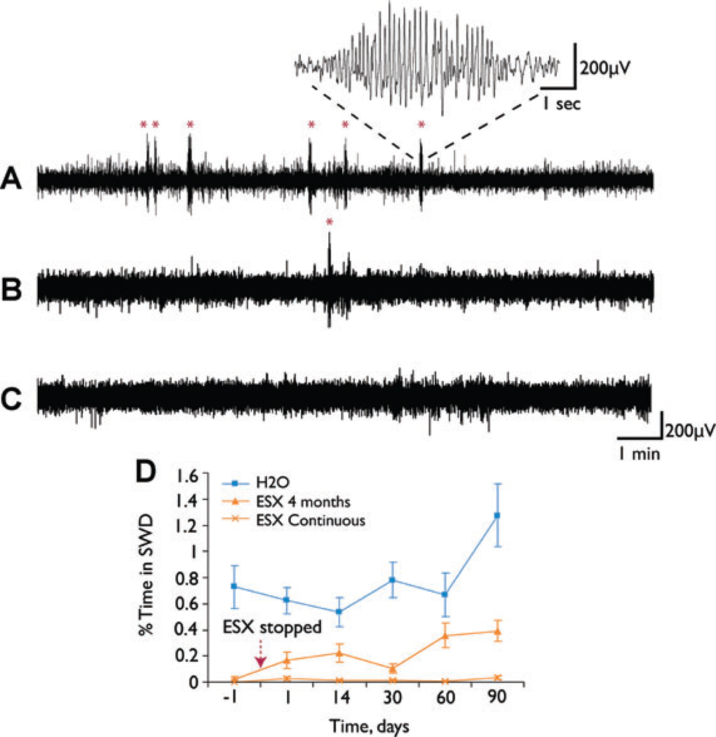
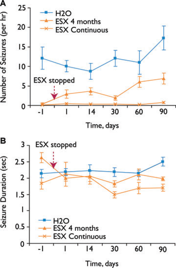
Methods: We used Wistar albino Glaxo rats of Rijswijk (WAG/Rij) rats, an established model of human absence epilepsy. Oral ethosuximide was given from age p21 to 5 months, covering the usual period in which seizures develop in this model (age approximately 3 months). Two experiments were performed: (1) cortical expression of ion channels Nav1.1, Nav1.6, and HCN1 (previously shown to be dysregulated in WAG/Rij) measured by immunocytochemistry in adult treated rats; and (2) electroencephalogram (EEG) recordings to measure seizure severity at serial time points after stopping the treatment.

Results: Early treatment with ethosuximide blocked changes in the expression of ion channels Nav1.1, Nav1.6, and HCN1 normally associated with epilepsy in this model. In addition, the treatment led to a persistent suppression of seizures, even after therapy was discontinued. Thus, animals treated with ethosuximide from age p21 to 5 months still had a marked suppression of seizures at age 8 months.

Discussion: These findings suggest that early treatment during development may provide a new strategy for preventing epilepsy in susceptible individuals. If confirmed with other drugs and epilepsy paradigms, the availability of a model in which epileptogenesis can be controlled has important implications both for future basic studies, and human therapeutic trials.


Figures

Figure 1
Figure 1
Experimental design for measuring effects of early ethosuximide treatment on ion channel expression (A) and epilepsy (B). (A) Experiment 1: Epileptic WAG/Rij rats and nonepileptic Wistar control rats (NE) were given either normal drinking water (H2O), or ethosuximide (ESX) 300 mg/kg/day from age p21 through age 5 months. EEG was recorded at age 5 months to determine amount of SWD. Animals were then sacrificed for immunocytochemistry to determine expression levels of Nav1.1, Nav1.6, and HCN1 in layer II–III cortical somatosensory neurons, shown previously to have abnormal protein expression levels in epileptic WAG/Rij rats (Klein et al., 2004; Strauss et al., 2004). Number of rats in each group were: NE control H2O (n = 8), NE control ESX (n = 8), WAG/Rij H2O (n = 7), and WAG/Rij ESX (n = 8). (B) Experiment 2: Epileptic WAG/Rij rats were given either normal drinking water (H2O group, n = 13); ethosuximide 300 mg/kg/day from age p21 through age 5 months and then normal drinking water from age 5 to 8 months (ESX 4 month group, n = 11); or ethosuximide 300 mg/kg/day continuously from age p21 through 8 months (ESX continuous group, n = 13). EEG was recorded in the ESX 4 month group, and in age-matched animals from the other two groups 1 day before stopping ethosuximide, and 1, 14, 30, 60, and 90 days after stopping ethosuximide. Epilepsia © ILAE
Figure 2
Figure 2
Examples of EEG recordings from Experiment 1 (Fig. 1A). Oral ethosuximide blocks SWD in WAG/Rij rats. (A) Recording from WAG/Rij rat on normal drinking water at age 5 months. Several episodes of typical SWD were observed (marked by *), with rhythmic large amplitude 7–8 Hz spike and wave lasting for a few seconds. Inset shows time expansion of one episode. (B) Recording from age-matched WAG/Rij rat on ethosuximide 300 mg/kg/day shows complete blockade of SWD episodes. Recordings from age-matched nonepileptic (NE) control rats on normal drinking water (C) or on ethosuximide 300 mg/kg/day (D) likewise showed a lack of SWD episodes. Epilepsia © ILAE
Figure 3
Figure 3
Early ethosuximide treatment blocks changes in ion channel expression (Experiment 1). (A–L) Examples of immunocytochemistry for Nav1.1 (A–D), Nav1.6 (E–H), and HCN1 (I–L) in somatosensory cortex layers II–III. (A–D) Nav1.1 is increased in WAG/Rij rats on normal drinking water (H2O) (C). The increase in Nav1.1 is blocked in WAG/Rij rats treated with early ethosuximide (ESX) (D). Nav1.1 expression in treated animals (D) resembles nonepileptic (NE) control animals (A, B). (E–H) Nav1.6 is increased in WAG/Rij rats on H2O (G). The increase in Nav1.6 is blocked by early ESX treatment (H), producing levels that resemble NE controls (E, F). (I–L) HCN1 is decreased in WAG/Rij rats on H2O (K). The decrease in HCN1 is blocked by early ESX treatment (L), producing levels that resemble NE controls (I, J). (M) Quantitative optical density changes in immunocytochemistry for Nav1.1, Nav1.6, and HCN1 in somatosensory cortical layer II–III neurons. Epileptic WAG/Rij rats on H2O have increased expression of Nav1.1 and Nav1.6, and decreased expression of HCN1. Expression levels for these channels were significantly different in the WAG/Rij H2O group compared to all other groups (WAG/Rij H2O group vs. NE H2O, NE ESX and WAG/Rij ESX, respectively, F = 36.75, 38.62, 44.32; p = 1.07 × 10⁻⁹, 6.36 × 10⁻¹⁰, 9.09 × 10⁻¹¹, d.f. = 2, between groups ANOVA for all three channels; *p < 0.01, post hoc between-group comparisons for each individual channel, Bonferroni corrected), and the other three groups did not differ significantly from each other (F = 0.24, p = 0.79, d.f. = 2). Images shown in A–L were equivalently enhanced (i.e., the identical brightness and contrast enhancements were made to each picture) to help demonstrate the differences in a way that would be clear in a printed format. For quantification (M), raw unenhanced images were used. Scale bar (K, applies for all panels) = 50 µm. Epilepsia © ILAE
Figure 4
Figure 4
Early ethosuximide (ESX) treatment persistently suppresses the development of SWD (Experiment 2). (A–C) Examples of EEG recordings from WAG/Rij rats. (A) WAG/Rij rat at age 8 months, on normal drinking water throughout development, has frequent SWD (marked by *). Inset shows typical SWD at higher gain. (B) WAG/Rij rat at age 8 months, treated with 300 mg/kg/day ESX in drinking water from p21 through age 5 months, and normal water from age 5 through 8 months (ESX 4 month group). SWD remain suppressed 90 days after stopping treatment (age 8 months). (C) WAG/Rij rat at age 8 months, treated with ESX 300 mg/kg/day from p21 onward (ESX continuous group), shows complete blockade of SWD. (D) Quantification of effects of early ESX treatment on percent time in SWD = (total SWD time/total recording time) × 100%. WAG/Rij rats were given either normal drinking water (■), ethosuximide 300 mg/kg/day from p21 through age 5 months (ESX 4 month group) (▲), or ethosuximide 300 mg/kg/day from p21 onward (ESX continuous group) (X). Recordings were done just before, and at serial time points after the ethosuximide was stopped in the ESX 4 month group, and in age-matched controls (see Fig. 1B). Even after stopping ESX, percent time in SWD remained markedly reduced in the treated WAG/Rij rats (ESX 4 months group) when comparing all time points for days 1 through 90 to WAG/Rij rats on normal H2O (F = 38.18, p = 4.32 × 10⁻⁹, d.f. = 2, between groups MANOVA; p < 0.0002, post hoc Games–Howell test for ESX 4 months vs. H2O groups). Epilepsia © ILAE
Figure 5
Figure 5
Early ethosuximide treatment persistently suppresses the number but not the duration of SWD episodes. (A) Effects of early ESX treatment on number of SWD episodes per hour. WAG/Rij rats were given either normal drinking water (■), ethosuximide 300 mg/kg/day from p21 through age 5 months (ESX 4 month group) (▲), or ethosuximide 300 mg/kg/day from p21 onward (ESX continuous group) (X). Recordings were done just before, and at serial time points after the ethosuximide was stopped in the ESX 4 month group, and in age-matched controls (see Fig. 1B). Even after stopping ESX, the number of SWD per hour remained markedly reduced in the treated WAG/Rij rats (ESX 4 months group) when comparing all time points for days 1 through 90 to WAG/Rij rats on normal H2O (F = 42.82, p = 1.20 × 10⁻⁹, d.f. = 2, between groups MANOVA; p = 0.0001, post hoc Games–Howell test for ESX 4 months vs. H2O groups). (B) Effects of early ESX treatment on SWD duration. Experimental groups and time points were the same as in (A). Treatment had no significant effect on SWD duration (F = 2.47, p = 0.14, MANOVA). Animals and EEG data for (A) and (B) were the same as in Fig. 4D. Epilepsia © ILAE

References

    1. Avoli M, Rogawski MA, Avanzini G. Generalized epileptic disorders: an update. Epilepsia. 2001;42:445–457. - PubMed
    1. Bartolomei F, Gastaldi M, Massacrier A, Planells R, Nicolas S, Cau P. Changes in the mRNAs encoding subtypes I, II and III sodium channel alpha subunits following kainate-induced seizures in rat brain. J Neurocytol. 1997;26:667–678. - PubMed
    1. Ben-Ari Y, Holmes GL. Effects of seizures on developmental processes in the immature brain. Lancet Neurol. 2006;5:1055–1063. - PubMed
    1. Bender RA, Soleymani SV, Brewster AL, Nguyen ST, Beck H, Mathern GW, Baram TZ. Enhanced expression of a specific hyperpolarization-activated cyclic nucleotide-gated cation channel (HCN) in surviving dentate gyrus granule cells of human and experimental epileptic hippocampus. J Neurosci. 2003;23:6826–6836. - PMC - PubMed
    1. Berkovic SF, Mulley JC, Scheffer IE, Petrou S. Human epilepsies: interaction of genetic and acquired factors. Trends Neurosci. 2006;29:391–397. - PubMed

Publication types

MeSH terms