. 2008 Feb;86(2):226-34.
doi: 10.1016/j.exer.2007.10.012. Epub 2007 Nov 5.

Protein kinase C epsilon activates lens mitochondrial cytochrome c oxidase subunit IV during hypoxia

Affiliations

Protein kinase C epsilon activates lens mitochondrial cytochrome c oxidase subunit IV during hypoxia

Michael Barnett et al. Exp Eye Res. 2008 Feb.

Abstract

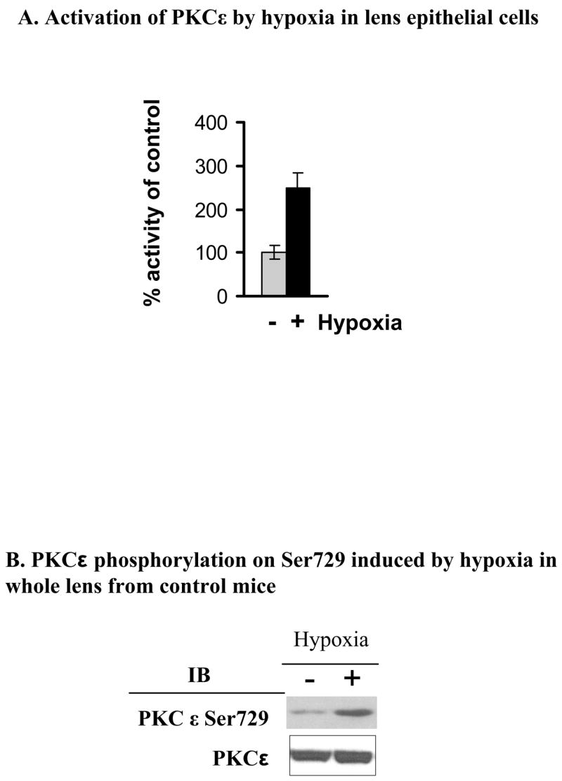
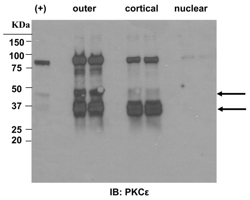
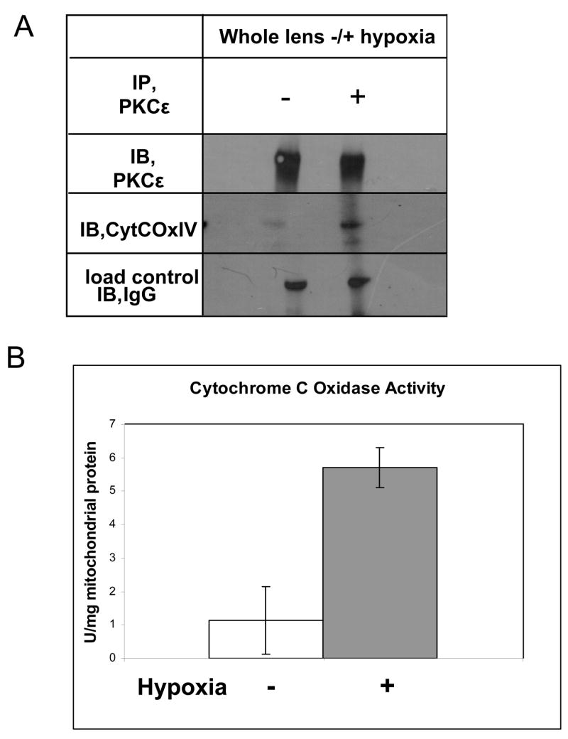
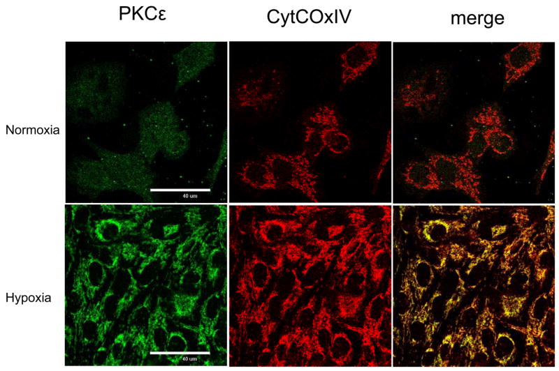
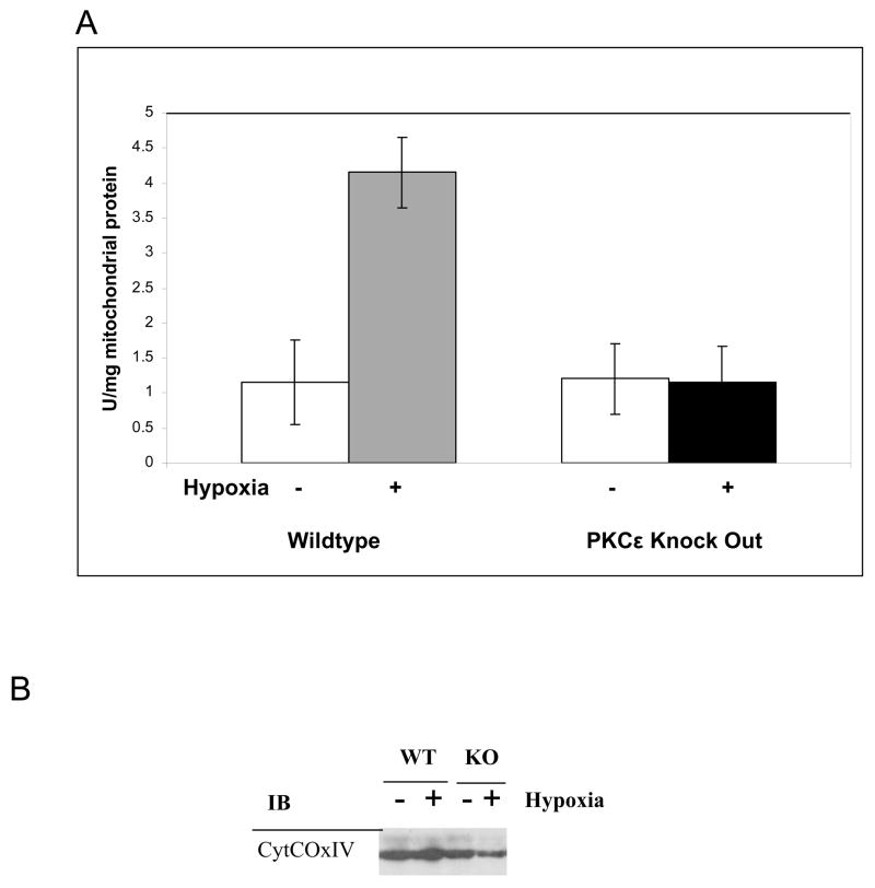
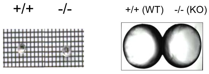
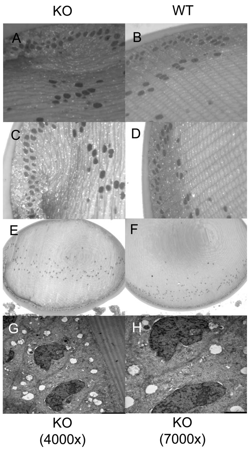
Protein kinase C (PKC) isoforms have been identified as major cellular signaling proteins that act directly in response to oxidation conditions. In retina and lens two isoforms of PKC respond to changes in oxidative stress, PKCgamma and PKCepsilon, while only PKCepsilon is found in heart. In heart the PKCepsilon acts on connexin 43 to protect from hypoxia. The presence of both isoforms in the lens led to this study to determine if lens PKCepsilon had unique targets. Both lens epithelial cells in culture and whole mouse lens were examined using PKC isoform-specific enzyme activity assays, co-immunoprecipitation, confocal microscopy, immunoblots, and light and electron microscopy. PKCepsilon was found in lens epithelium and cortex but not in the nucleus of mouse lens. The PKCepsilon isoform was activated in both epithelium and whole lens by 5% oxygen when compared to activity at 21% oxygen. In hypoxic conditions (5% oxygen) the PKCepsilon co-immunoprecipitated with the mitochondrial cytochrome c oxidase IV subunit (CytCOx). Concomitant with this the CytCOx enzyme activity was elevated and increased co-localization of CytCOx with PCKvarepsilon was observed using immunolabeling and confocal microscopy. In contrast, no hypoxia-induced activation of CytCOx was observed in lenses from the PKCepsilon knockout mice. Lens from 6-week-old PKCepsilon knockout mice had a disorganized bow region which was filled with vacuoles indicating a possible loss of mitochondria but the size of the lens was not altered. Electron microscopy demonstrated that the nuclei of the PCKepsilon knockout mice were abnormal in shape. Thus, PKCepsilon is found to be activated by hypoxia and this results in the activation of the mitochondrial protein CytCOx. This could protect the lens from mitochondrial damage under the naturally hypoxic conditions observed in this tissue. Lens oxygen levels must remain low. Elevation of oxygen which occurs during vitreal detachment or liquification is associated with cataracts. We hypothesize that elevated oxygen could cause inhibition of PKCepsilon resulting in a loss of mitochondrial protection.


Figures

Figure 1
Figure 1. Hypoxia-induced PKCε activation in the lens
A. Enzyme activity of PKCε in N/N1003A rabbit lens epithelial cells was determined as described in the Methods Section. In the wild type lens epithelial cells (N/N1003A) treated with hypoxia (5% O2) or normoxic (21 % O2) for 12 hours, PKCε was activated about 2.5 fold over control (set at 100%). Equal PKCε per lane was verified using PKCε antisera and western blots. B. The whole lenses from control mice were made hypoxic as described. PKCε phosphorylation on Ser729 was determined by Western blotting using anti-phospho-PKCε Ser729 antisera. Total PKCε is a loading control.
Figure 2
Figure 2. PKCε is widely expressed in the lens epithelium and cortex
Lenses of control mice were dissected as described. Tissue homogenates were subjected to Western blotting to detect PKCε expression. += PKCε positive control included with antisera. Lanes are duplicates. Arrows indicate the catalytic fragments of PKCε.
Figure 3
Figure 3. Analysis of Cytochrome C Oxidase IV interaction with PKCε and activity of CytCox in lenses treated with or without hypoxia
A. Cell lysates from the control mouse lenses with or without hypoxia were used to co-immunoprecipitate PKCε and its associated protein CytCoxIV. The interaction of PKCε with CytCoxIV is sharply enhanced from hypoxia. B. Cytochrome C Oxidase activity measurements show that the enzyme is activated up to six fold in mouse lenses treated with hypoxia for two hours. IP= immunoprecipitation antisera, IB=immunoblot antisera, +/− (Hypoxia).
Figure 4
Figure 4. Co-localization of PKCε with CytCoxIV after hypoxia
N/N cells were seeded in 12 well plates to 70% confluence before hypoxia treatment at 5% O2 for 12 hr. Cells were fixed with 2.5% paraformaldehyde for 5 min., blocked with 3% BSA in PBS for 1 hr at room temperature, and then labeled with anti-PKCε (Abcom, Cambridge MA) or CytCOxIV (MitoSciences, Eugene, OR) for 2 hr at room temperature. After washing, the cells were incubated with the secondary antisera Alexa Fluor 568 goat anti-rabbit IgG (H+L) or Alexa Fluor 488 goat anti-mouse IgG (H+L) (Molecular Probes, Oregon). The cells were then mounted on slides and examined using a Nikon C1 scanning confocal microscope.
Figure 5
Figure 5. PKCε and its associated protein CytCoxIV in the control and PKCε knockout mice
Proteins of whole lens homogenates (6 weeks old) were resolved by SDS-PAGE and Western blotted using specific antisera as shown. The PKCε knockout mice used in these experiments do not contain detectable levels of PKCε (A) but do contain comparable amounts of CytCOxIV (B). N/N 1003A cell lysates were loaded in left lane. Lanes are duplicates. Upper bands are nonspecific reactions.
Figure 6
Figure 6. Cytochrome C Oxidase activation in the lenses of control but not PKCε knockout mice with and without hypoxia
A. Cytochrome C oxidase enzyme activity is not increased by hypoxia in PKCε knockout mice. B. Total CytCOxIV levels in the PKCε knockout and control mice with or without hypoxia. KO, PKCε knockout; WT, wild type control mouse. All mice used at 6 weeks of age.
Figure 7
Figure 7. Wild type and PKCε knockout mouse lenses are not altered in size
Shown are pictures of fresh mouse lens (6 week old) from WT or KO mice. A. Grid picture. B. Enlarged.
Figure 8
Figure 8. Structural alteration of the PKCε knockout mouse lens
A-F. Lenses from PKCε knockout mice (KO) (A, C, and E) were compared to the control mouse lenses (WT) (B, D, and F) (6 week old). Whole lenses are pictured in E and F, at 10X. Light microscopy images of stained sections were taken at 40X (A-D). Nuclear disorganization and large numerous vacuoles were observed in the knockout lenses but not the wild type lenses. G-H. Electron microscopy images of PKCε knockout lenses at 4000X (G) or 7000X (H) show large vacuoles, distorted nuclei, and lack of mitochondria. KO, PKCε knockout; WT, wild type control mouse

References

    1. Abeliovich A, Paylor R, Chen C, Kim JJ, Wehner JM, Tonegawa S. PKCgamma mutant mice exhibit mild deficients in spatial and contextual learning. Cell. 1993;75:1263–1271. - PubMed
    1. Abeliovich A, Chen C, Goda Y, Silva AJ, Stevens CF, Tonegawa S. Modified hippocampal long-term potentiation in PKC γ-mutant mice. Cell. 1993;75:1253–1262. - PubMed
    1. Baines CP, Zhang J, Wang GW, Zheng YT, Xiu JX, Cardwell EM, Bolli R, Ping P. Mitochondrial PKCepsilon and MAPK form signaling modules in the murine heart: enhanced mitochondrial PKCepsilon-MAPK interactions and differential MAPK activation in PKCepsilon-induced cardioprotection. Circ Res. 2002;90:390–397. - PubMed
    1. Baines CP, Song CX, Zheng YT, Wang GW, Zhang J, Wang OL, Guo Y, Bolli R, Cardwell EM, Ping P. Protein kinase Cepsilon interacts with and inhibits the permeability transition pore in cardiac mitochondria. Circ Res. 2003;92:873–880. - PMC - PubMed
    1. Barnett ME, Madgwick DK, Takemoto DJ. Protein kinase C as a stress sensor. Cell Signal. 2007;19:1820–1829. - PMC - PubMed

Publication types

Substances